3JQQ

Crystal structure of the H286K mutant of Ferredoxin-NADP+ reductase from Plasmodium falciparum in complex with 2'P-AMP


Experimental Data Snapshot

  • Method: X-RAY DIFFRACTION
  • Resolution: 2.20 Å
  • R-Value Free: 0.308 
  • R-Value Work: 0.256 
  • R-Value Observed: 0.259 

wwPDB Validation   3D Report Full Report


Ligand Structure Quality Assessment 


This is version 1.3 of the entry. See complete history


Literature

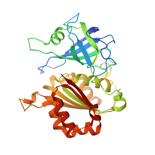
Plasmodium falciparum ferredoxin-NADP+ reductase His286 plays a dual role in NADP(H) binding and catalysis

Crobu, D.Canevari, G.Milani, M.Pandini, V.Vanoni, M.A.Bolognesi, M.Zanetti, G.Aliverti, A.

(2009) Biochemistry 48: 9525-9533

  • DOI: https://doi.org/10.1021/bi9013209
  • Primary Citation of Related Structures:  
    3JQP, 3JQQ, 3JQR

  • PubMed Abstract: 

    The NADP-binding site of Plasmodium falciparum ferredoxin-NADP(+) reductase contains two basic residues, His286 and Lys249, conserved within the Plasmodium genus, but not in other plant-type homologues. Previous crystal studies indicated that His286 interacts with the adenine ring and with the 5'-phosphate of 2'-P-AMP, a ligand that mimics the adenylate moiety of NADP(H). Here we show that replacement of His286 with aliphatic residues results both in a decrease in the affinity of the enzyme for NADPH and in a decrease in k(cat), due to a lowered hydride-transfer rate. Unexpectedly, the mutation to Gln produces an enzyme more active than the wild-type one, whereas the change to Lys destabilizes the nicotinamide-isoalloxazine interaction, decreasing k(cat). On the basis of the crystal structure of selected mutants complexed with 2'-P-AMP, we conclude that the His286 side chain plays a dual role in catalysis both by providing binding energy for NADPH and by favoring the catalytically competent orientation of its nicotinamide ring. For the latter function, the H-bonding potential rather than the positively charged state of the His286 imidazole seems sufficient. Furthermore, we show that the Lys249Ala mutation decreases K(m)(NADPH) and K(d) for NADP(+) or 2'-P-AMP by a factor of 10. We propose that the Lys249 side chain participates in substrate recognition by interacting with the 2'-phosphate of NADP(H) and that this interaction was not observed in the crystal form of the enzyme-2'-P-AMP complex due to a conformational perturbation of the substrate-binding loop induced by dimerization.


  • Organizational Affiliation

    Dipartimento di Scienze Biomolecolari e Biotecnologie, Universita degli Studi di Milano, via Celoria 26, 20133 Milano, Italy.


Macromolecules
Find similar proteins by:  (by identity cutoff)  |  3D Structure
Entity ID: 1
MoleculeChains Sequence LengthOrganismDetailsImage
Ferredoxin NADP reductase
A, B, C, D, E
A, B, C, D, E, F
316Plasmodium falciparumMutation(s): 1 
Gene Names: PfFNRPFF1115w
EC: 1.18.1.2
UniProt
Find proteins for C6KT68 (Plasmodium falciparum (isolate 3D7))
Explore C6KT68 
Go to UniProtKB:  C6KT68
Entity Groups  
Sequence Clusters30% Identity50% Identity70% Identity90% Identity95% Identity100% Identity
UniProt GroupC6KT68
Sequence Annotations
Expand
  • Reference Sequence
Small Molecules
Ligands 2 Unique
IDChains Name / Formula / InChI Key2D Diagram3D Interactions
FAD
Query on FAD

Download Ideal Coordinates CCD File 
G [auth A]
I [auth B]
K [auth C]
M [auth D]
O [auth E]
G [auth A],
I [auth B],
K [auth C],
M [auth D],
O [auth E],
Q [auth F]
FLAVIN-ADENINE DINUCLEOTIDE
C27 H33 N9 O15 P2
VWWQXMAJTJZDQX-UYBVJOGSSA-N
A2P
Query on A2P

Download Ideal Coordinates CCD File 
H [auth A]
J [auth B]
L [auth C]
N [auth D]
P [auth E]
H [auth A],
J [auth B],
L [auth C],
N [auth D],
P [auth E],
R [auth F]
ADENOSINE-2'-5'-DIPHOSPHATE
C10 H15 N5 O10 P2
AEOBEOJCBAYXBA-KQYNXXCUSA-N
Binding Affinity Annotations 
IDSourceBinding Affinity
A2P Binding MOAD:  3JQQ Ki: 2900 (nM) from 1 assay(s)
Experimental Data & Validation

Experimental Data

  • Method: X-RAY DIFFRACTION
  • Resolution: 2.20 Å
  • R-Value Free: 0.308 
  • R-Value Work: 0.256 
  • R-Value Observed: 0.259 
  • Space Group: P 31
Unit Cell:
Length ( Å )Angle ( ˚ )
a = 124.243α = 90
b = 124.243β = 90
c = 134.255γ = 120
Software Package:
Software NamePurpose
DNAdata collection
MOLREPphasing
REFMACrefinement
MOSFLMdata reduction
SCALAdata scaling

Structure Validation

View Full Validation Report


Ligand Structure Quality Assessment 


Entry History 

Deposition Data

Revision History  (Full details and data files)

  • Version 1.0: 2010-01-12
    Type: Initial release
  • Version 1.1: 2011-07-13
    Changes: Version format compliance
  • Version 1.2: 2021-11-10
    Changes: Database references, Derived calculations
  • Version 1.3: 2023-11-01
    Changes: Data collection, Refinement description