3JBZ

Crystal structure of mTOR docked into EM map of dimeric ATM kinase


Experimental Data Snapshot

  • Method: ELECTRON MICROSCOPY
  • Resolution: 28.0 Å
  • Aggregation State: PARTICLE 
  • Reconstruction Method: SINGLE PARTICLE 

wwPDB Validation   3D Report Full Report


This is version 1.4 of the entry. See complete history


Literature

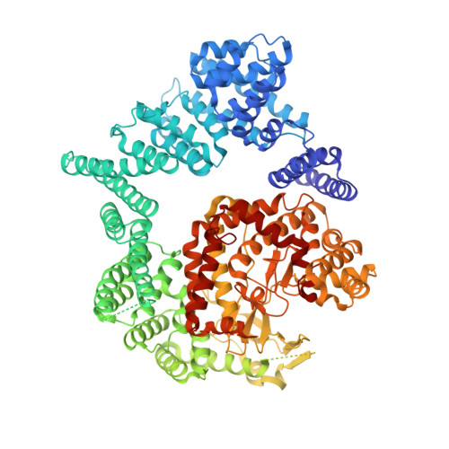
Structure of the human dimeric ATM kinase.

Lau, W.C.Li, Y.Liu, Z.Gao, Y.Zhang, Q.Huen, M.S.

(2016) Cell Cycle 15: 1117-1124

  • DOI: https://doi.org/10.1080/15384101.2016.1158362
  • Primary Citation of Related Structures:  
    3JBZ

  • PubMed Abstract: 

    DNA-double strand breaks activate the serine/threonine protein kinase ataxia-telangiectasia mutated (ATM) to initiate DNA damage signal transduction. This activation process involves autophosphorylation and dissociation of inert ATM dimers into monomers that are catalytically active. Using single-particle electron microscopy (EM), we determined the structure of dimeric ATM in its resting state. The EM map could accommodate the crystal structure of the N-terminal truncated mammalian target of rapamycin (mTOR), a closely related enzyme of the phosphatidylinositol 3-kinase-related protein kinase (PIKK) family, allowing for the localization of the N- and the C-terminal regions of ATM. In the dimeric structure, the actives sites are buried, restricting the access of the substrates to these sites. The unanticipated domain organization of ATM provides a basis for understanding its mechanism of inhibition.


  • Organizational Affiliation

    a School of Biomedical Sciences, The University of Hong Kong , Hong Kong.


Macromolecules
Find similar proteins by:  (by identity cutoff)  |  3D Structure
Entity ID: 1
MoleculeChains Sequence LengthOrganismDetailsImage
Serine/threonine-protein kinase mTOR1,165Homo sapiensMutation(s): 0 
Gene Names: FRAPFRAP1FRAP2MTORRAFT1RAPT1
EC: 2.7.11.1
UniProt & NIH Common Fund Data Resources
Find proteins for P42345 (Homo sapiens)
Explore P42345 
Go to UniProtKB:  P42345
PHAROS:  P42345
GTEx:  ENSG00000198793 
Entity Groups  
Sequence Clusters30% Identity50% Identity70% Identity90% Identity95% Identity100% Identity
UniProt GroupP42345
Sequence Annotations
Expand
  • Reference Sequence
Experimental Data & Validation

Experimental Data

  • Method: ELECTRON MICROSCOPY
  • Resolution: 28.0 Å
  • Aggregation State: PARTICLE 
  • Reconstruction Method: SINGLE PARTICLE 
EM Software:
TaskSoftware PackageVersion
RECONSTRUCTIONSIMPLE
RECONSTRUCTIONEMAN2

Structure Validation

View Full Validation Report


Entry History 

Deposition Data

  • Released Date: 2016-11-16 
  • Deposition Author(s): Lau, W.C.Y.

Revision History  (Full details and data files)

  • Version 1.0: 2016-11-16
    Type: Initial release
  • Version 1.1: 2016-11-23
    Changes: Source and taxonomy
  • Version 1.2: 2019-10-09
    Changes: Data collection, Database references, Other
  • Version 1.3: 2019-12-18
    Changes: Data collection
  • Version 1.4: 2024-03-20
    Changes: Data collection, Database references, Derived calculations, Refinement description