Three-dimensional electron crystallography of protein microcrystals.
Shi, D., Nannenga, B.L., Iadanza, M.G., Gonen, T.(2013) Elife 2: e01345-e01345
- PubMed: 24252878 Search on PubMedSearch on PubMed Central
- DOI: https://doi.org/10.7554/eLife.01345
- Primary Citation Related Structures:
3J4G - PubMed Abstract:
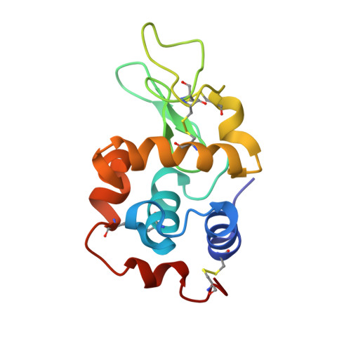
We demonstrate that it is feasible to determine high-resolution protein structures by electron crystallography of three-dimensional crystals in an electron cryo-microscope (CryoEM). Lysozyme microcrystals were frozen on an electron microscopy grid, and electron diffraction data collected to 1.7 Å resolution. We developed a data collection protocol to collect a full-tilt series in electron diffraction to atomic resolution. A single tilt series contains up to 90 individual diffraction patterns collected from a single crystal with tilt angle increment of 0.1-1° and a total accumulated electron dose less than 10 electrons per angstrom squared. We indexed the data from three crystals and used them for structure determination of lysozyme by molecular replacement followed by crystallographic refinement to 2.9 Å resolution. This proof of principle paves the way for the implementation of a new technique, which we name 'MicroED', that may have wide applicability in structural biology. DOI: http://dx.doi.org/10.7554/eLife.01345.001.
- Janelia Farm Research Campus, Howard Hughes Medical Institute, Ashburn, United States.
Organizational Affiliation: