3IOR | pdb_00003ior

Huntingtin amino-terminal region with 17 Gln residues - crystal C95


Experimental Data Snapshot

  • Method: X-RAY DIFFRACTION
  • Resolution: 3.60 Å
  • R-Value Free: 
    0.267 (Depositor), 0.222 (DCC) 
  • R-Value Work: 
    0.258 (Depositor) 
  • R-Value Observed: 
    0.258 (Depositor) 

wwPDB Validation 3D Report Full Report

Validation slider image for 3IOR

This is version 1.4 of the entry. See complete history

Literature

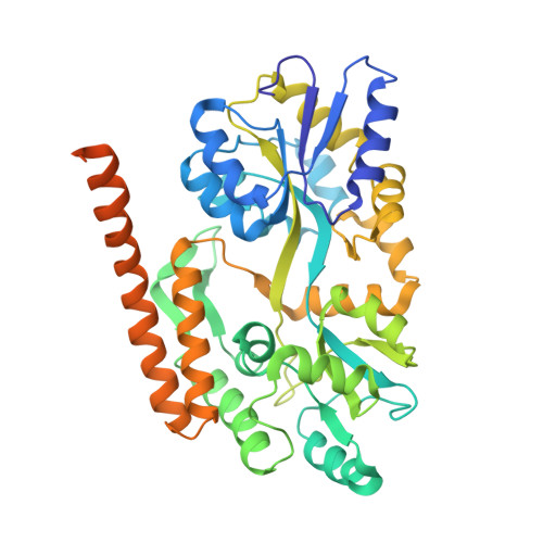
Secondary structure of Huntingtin amino-terminal region.

Kim, M.W.Chelliah, Y.Kim, S.W.Otwinowski, Z.Bezprozvanny, I.

(2009) Structure 17: 1205-1212

  • DOI: https://doi.org/10.1016/j.str.2009.08.002
  • Primary Citation Related Structures: 
    3IO4, 3IO6, 3IOR, 3IOT, 3IOU, 3IOV, 3IOW

  • PubMed Abstract: 

    Huntington's disease is a genetic neurodegenerative disorder resulting from polyglutamine (polyQ) expansion (>36Q) within the first exon of Huntingtin (Htt) protein. We applied X-ray crystallography to determine the secondary structure of the first exon (EX1) of Htt17Q. The structure of Htt17Q-EX1 consists of an amino-terminal alpha helix, poly17Q region, and polyproline helix formed by the proline-rich region. The poly17Q region adopts multiple conformations in the structure, including alpha helix, random coil, and extended loop. The conformation of the poly17Q region is influenced by the conformation of neighboring protein regions, demonstrating the importance of the native protein context. We propose that the conformational flexibility of the polyQ region observed in our structure is a common characteristic of many amyloidogenic proteins. We further propose that the pathogenic polyQ expansion in the Htt protein increases the length of the random coil, which promotes aggregation and facilitates abnormal interactions with other proteins in cells.


  • Organizational Affiliation
    • Department of Biochemistry, University of Texas Southwestern Medical Center at Dallas, Dallas, TX 75390, USA. MeeWhi.Kim@UTSouthwestern.edu

Macromolecule Content 

  • Total Structure Weight: 148.58 kDa 
  • Atom Count: 9,166 
  • Modeled Residue Count: 1,202 
  • Deposited Residue Count: 1,347 
  • Unique protein chains: 1

Macromolecules

Find similar proteins by:|  3D Structure
Entity ID: 1
MoleculeChains  Sequence LengthOrganismDetailsImage
Maltose-binding protein, huntingtin fusion protein
A, B, C
449Escherichia coli K-12Homo sapiens
This entity is chimeric
Mutation(s): 0 
Gene Names: malEb4034
UniProt & NIH Common Fund Data Resources
Find proteins for P0AEX9 (Escherichia coli (strain K12))
Explore P0AEX9 
Go to UniProtKB:  P0AEX9
Find proteins for P42858 (Homo sapiens)
Explore P42858 
Go to UniProtKB:  P42858
PHAROS:  P42858
GTEx:  ENSG00000197386 
Entity Groups
Sequence Clusters30% Identity50% Identity70% Identity90% Identity95% Identity100% Identity
UniProt GroupsP42858P0AEX9
Sequence Annotations
Expand
Reference Sequence

Small Molecules

Ligands 2 Unique
IDChains Name / Formula / InChI Key2D Diagram3D Interactions
ZN

Query on ZN


Download:Ideal Coordinates CCD File
D [auth A]
E [auth A]
G [auth B]
H [auth B]
I [auth B]
D [auth A],
E [auth A],
G [auth B],
H [auth B],
I [auth B],
L [auth C]
ZINC ION
Zn
PTFCDOFLOPIGGS-UHFFFAOYSA-N
CA

Query on CA


Download:Ideal Coordinates CCD File
F [auth A]
J [auth B]
K [auth B]
M [auth C]
N [auth C]
F [auth A],
J [auth B],
K [auth B],
M [auth C],
N [auth C],
O [auth C]
CALCIUM ION
Ca
BHPQYMZQTOCNFJ-UHFFFAOYSA-N

Experimental Data & Validation

Experimental Data

  • Method: X-RAY DIFFRACTION
  • Resolution: 3.60 Å
  • R-Value Free:  0.267 (Depositor), 0.222 (DCC) 
  • R-Value Work:  0.258 (Depositor) 
  • R-Value Observed: 0.258 (Depositor) 
Space Group: C 1 2 1
Unit Cell:
Length ( Å )Angle ( ˚ )
a = 163.08α = 90
b = 100.98β = 95.14
c = 137.86γ = 90
Software Package:
Software NamePurpose
HKL-2000data collection
PHASERphasing
REFMACrefinement
HKL-2000data reduction
HKL-2000data scaling

Structure Validation

View Full Validation Report


Entry History 

Deposition Data

  • Released Date: 2009-09-29 
  • Deposition Author(s): Kim, M.W.

Revision History  (Full details and data files)

  • Version 1.0: 2009-09-29
    Type: Initial release
  • Version 1.1: 2011-07-13
    Changes: Version format compliance
  • Version 1.2: 2015-06-24
    Changes: Database references, Source and taxonomy
  • Version 1.3: 2017-06-28
    Changes: Database references, Structure summary
  • Version 1.4: 2024-02-21
    Changes: Data collection, Database references, Derived calculations