3IGG | pdb_00003igg

Novel CDK-5 inhibitors - crystal structure of inhibitor EFQ with CDK-2


Experimental Data Snapshot

  • Method: X-RAY DIFFRACTION
  • Resolution: 1.80 Å
  • R-Value Free: 
    0.257 (Depositor), 0.240 (DCC) 
  • R-Value Work: 
    0.209 (Depositor), 0.199 (DCC) 
  • R-Value Observed: 
    0.209 (Depositor) 

wwPDB Validation 3D Report Full Report

Validation slider image for 3IGG

Ligand Structure Quality Assessment 


This is version 1.2 of the entry. See complete history

Literature

Potent and cellularly active 4-aminoimidazole inhibitors of cyclin-dependent kinase 5/p25 for the treatment of Alzheimer's disease.

Helal, C.J.Kang, Z.Lucas, J.C.Gant, T.Ahlijanian, M.K.Schachter, J.B.Richter, K.E.Cook, J.M.Menniti, F.S.Kelly, K.Mente, S.Pandit, J.Hosea, N.

(2009) Bioorg Med Chem Lett 19: 5703-5707

  • DOI: https://doi.org/10.1016/j.bmcl.2009.08.019
  • Primary Citation Related Structures: 
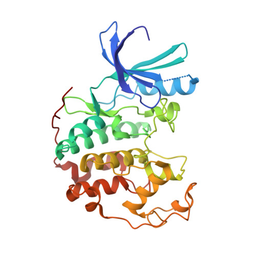
    3IG7, 3IGG

  • PubMed Abstract: 

    Utilizing structure-based drug design, a 4-aminoimidazole heterocyclic core was synthesized as a replacement for a 2-aminothiazole due to potential metabolically mediated toxicity. The synthetic route utilized allowed for ready synthesis of 1-substituted-4-aminoimidazoles. SAR exploration resulted in the identification of a novel cis-substituted cyclobutyl group that gave improved enzyme and cellular potency against cdk5/p25 with up to 30-fold selectivity over cdk2/cyclin E.


  • Organizational Affiliation
    • Neuroscience Medicinal Chemistry, Pfizer Global Research and Development, Eastern Point Road, Groton, CT 06340, USA. chris.j.helal@pfizer.com

Macromolecule Content 

  • Total Structure Weight: 34.3 kDa 
  • Atom Count: 2,479 
  • Modeled Residue Count: 291 
  • Deposited Residue Count: 299 
  • Unique protein chains: 1

Macromolecules

Find similar proteins by:|  3D Structure
Entity ID: 1
MoleculeChains  Sequence LengthOrganismDetailsImage
Cell division protein kinase 2299Homo sapiensMutation(s): 0 
Gene Names: CDK2
EC: 2.7.11.22
UniProt & NIH Common Fund Data Resources
Find proteins for P24941 (Homo sapiens)
Explore P24941 
Go to UniProtKB:  P24941
PHAROS:  P24941
GTEx:  ENSG00000123374 
Entity Groups
Sequence Clusters30% Identity50% Identity70% Identity90% Identity95% Identity100% Identity
UniProt GroupP24941
Sequence Annotations
Expand
Reference Sequence

Small Molecules

Ligands 1 Unique
IDChains Name / Formula / InChI Key2D Diagram3D Interactions
EFQ

Query on EFQ


Download:Ideal Coordinates CCD File
B [auth A]N-[1-(cis-3-hydroxycyclobutyl)-1H-imidazol-4-yl]-2-(4-methoxyphenyl)acetamide
C16 H19 N3 O3
SVPUOZWGAJVQMQ-BETUJISGSA-N

Experimental Data & Validation

Experimental Data

  • Method: X-RAY DIFFRACTION
  • Resolution: 1.80 Å
  • R-Value Free:  0.257 (Depositor), 0.240 (DCC) 
  • R-Value Work:  0.209 (Depositor), 0.199 (DCC) 
  • R-Value Observed: 0.209 (Depositor) 
Space Group: P 21 21 21
Unit Cell:
Length ( Å )Angle ( ˚ )
a = 53.47α = 90
b = 72.04β = 90
c = 72.04γ = 90
Software Package:
Software NamePurpose
REFMACrefinement
PDB_EXTRACTdata extraction

Structure Validation

View Full Validation Report


Ligand Structure Quality Assessment 


Entry History 

Deposition Data

  • Released Date: 2009-09-08 
  • Deposition Author(s): Pandit, J.

Revision History  (Full details and data files)

  • Version 1.0: 2009-09-08
    Type: Initial release
  • Version 1.1: 2011-07-13
    Changes: Version format compliance
  • Version 1.2: 2024-11-27
    Changes: Data collection, Database references, Derived calculations, Structure summary