3I80 | pdb_00003i80

Protein Tyrosine Phosphatase 1B - Transition state analog for the second catalytic step


Experimental Data Snapshot

  • Method: X-RAY DIFFRACTION
  • Resolution: 2.25 Å
  • R-Value Free: 
    0.235 (Depositor), 0.227 (DCC) 
  • R-Value Work: 
    0.199 (Depositor), 0.186 (DCC) 
  • R-Value Observed: 
    0.201 (Depositor) 

Starting Model: experimental
View more details

wwPDB Validation 3D Report Full Report

Validation slider image for 3I80

This is version 1.2 of the entry. See complete history

Literature

Insights into the reaction of protein-tyrosine phosphatase 1B: crystal structures for transition state analogs of both catalytic steps.

Brandao, T.A.Hengge, A.C.Johnson, S.J.

(2010) J Biological Chem 285: 15874-15883

  • DOI: https://doi.org/10.1074/jbc.M109.066951
  • Primary Citation Related Structures: 
    3I7Z, 3I80

  • PubMed Abstract: 

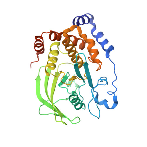
    Catalysis by protein-tyrosine phosphatase 1B (PTP1B) occurs through a two-step mechanism involving a phosphocysteine intermediate. We have solved crystal structures for the transition state analogs for both steps. Together with previously reported crystal structures of apo-PTP1B, the Michaelis complex of an inactive mutant, the phosphoenzyme intermediate, and the product complex, a full picture of all catalytic steps can now be depicted. The transition state analog for the first catalytic step comprises a ternary complex between the catalytic cysteine of PTP1B, vanadate, and the peptide DADEYL, a fragment of a physiological substrate. The equatorial vanadate oxygen atoms bind to the P-loop, and the apical positions are occupied by the peptide tyrosine oxygen and by the PTP1B cysteine sulfur atom. The vanadate assumes a trigonal bipyramidal geometry in both transition state analog structures, with very similar apical O-O distances, denoting similar transition states for both phosphoryl transfer steps. Detailed interactions between the flanking peptide and the enzyme are discussed.


  • Organizational Affiliation
    • Department of Chemistry and Biochemistry, Utah State University, Logan, Utah 84322-0300, USA.

Macromolecule Content 

  • Total Structure Weight: 37.88 kDa 
  • Atom Count: 2,701 
  • Modeled Residue Count: 298 
  • Deposited Residue Count: 321 
  • Unique protein chains: 1

Macromolecules

Find similar proteins by:|  3D Structure
Entity ID: 1
MoleculeChains  Sequence LengthOrganismDetailsImage
Tyrosine-protein phosphatase non-receptor type 1321Homo sapiensMutation(s): 0 
Gene Names: PTP1BPTPN1
EC: 3.1.3.48
UniProt & NIH Common Fund Data Resources
Find proteins for P18031 (Homo sapiens)
Explore P18031 
Go to UniProtKB:  P18031
PHAROS:  P18031
GTEx:  ENSG00000196396 
Entity Groups
Sequence Clusters30% Identity50% Identity70% Identity90% Identity95% Identity100% Identity
UniProt GroupP18031
Sequence Annotations
Expand
Reference Sequence

Experimental Data & Validation

Experimental Data

  • Method: X-RAY DIFFRACTION
  • Resolution: 2.25 Å
  • R-Value Free:  0.235 (Depositor), 0.227 (DCC) 
  • R-Value Work:  0.199 (Depositor), 0.186 (DCC) 
  • R-Value Observed: 0.201 (Depositor) 
Space Group: P 31 2 1
Unit Cell:
Length ( Å )Angle ( ˚ )
a = 87.84α = 90
b = 87.84β = 90
c = 103.79γ = 120
Software Package:
Software NamePurpose
d*TREKdata scaling
PHASERphasing
PHENIXrefinement
PDB_EXTRACTdata extraction
CrystalCleardata collection
d*TREKdata reduction

Structure Validation

View Full Validation Report


Entry History 

Deposition Data

Revision History  (Full details and data files)

  • Version 1.0: 2010-03-16
    Type: Initial release
  • Version 1.1: 2011-07-13
    Changes: Version format compliance
  • Version 1.2: 2023-09-06
    Changes: Data collection, Database references, Derived calculations, Refinement description