3I7G | pdb_00003i7g

MMP-13 in complex with a non zinc-chelating inhibitor


Experimental Data Snapshot

  • Method: X-RAY DIFFRACTION
  • Resolution: 1.95 Å
  • R-Value Free: 
    0.228 (Depositor), 0.221 (DCC) 
  • R-Value Work: 
    0.173 (Depositor), 0.168 (DCC) 
  • R-Value Observed: 
    0.176 (Depositor) 

wwPDB Validation 3D Report Full Report

Validation slider image for 3I7G

Ligand Structure Quality Assessment 


This is version 1.2 of the entry. See complete history

Literature

Improving potency and selectivity of a new class of non-Zn-chelating MMP-13 inhibitors.

Heim-Riether, A.Taylor, S.J.Liang, S.Gao, D.A.Xiong, Z.Michael August, E.Collins, B.K.Farmer, B.T.Haverty, K.Hill-Drzewi, M.Junker, H.D.Mariana Margarit, S.Moss, N.Neumann, T.Proudfoot, J.R.Keenan, L.S.Sekul, R.Zhang, Q.Li, J.Farrow, N.A.

(2009) Bioorg Med Chem Lett 19: 5321-5324

  • DOI: https://doi.org/10.1016/j.bmcl.2009.07.151
  • Primary Citation Related Structures: 
    3I7G, 3I7I

  • PubMed Abstract: 

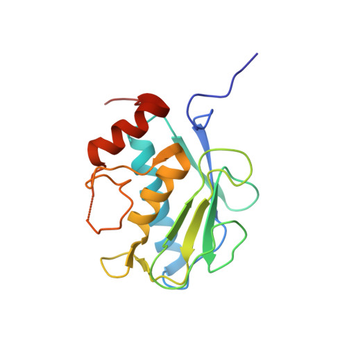
    Discovery and optimization of potency and selectivity of a non-Zn-chelating MMP-13 inhibitor with the aid of protein co-crystal structural information is reported. This inhibitor was observed to have a binding mode distinct from previously published MMP-13 inhibitors. Potency and selectivity were improved by extending the hit structure out from the active site into the S1' pocket.


  • Organizational Affiliation
    • Boehringer Ingelheim Pharmaceuticals, Department of Medicinal Chemistry, 900 Ridgebury Rd, Ridgefield, CT 06877, USA. alexander.heim-riether@boehringer-ingelheim.com

Macromolecule Content 

  • Total Structure Weight: 39.44 kDa 
  • Atom Count: 3,006 
  • Modeled Residue Count: 328 
  • Deposited Residue Count: 342 
  • Unique protein chains: 1

Macromolecules

Find similar proteins by:|  3D Structure
Entity ID: 1
MoleculeChains  Sequence LengthOrganismDetailsImage
Collagenase 3
A, B
171Homo sapiensMutation(s): 0 
Gene Names: MMP13
EC: 3.4.24
UniProt & NIH Common Fund Data Resources
Find proteins for P45452 (Homo sapiens)
Explore P45452 
Go to UniProtKB:  P45452
PHAROS:  P45452
GTEx:  ENSG00000137745 
Entity Groups
Sequence Clusters30% Identity50% Identity70% Identity90% Identity95% Identity100% Identity
UniProt GroupP45452
Sequence Annotations
Expand
Reference Sequence

Small Molecules

Ligands 4 Unique
IDChains Name / Formula / InChI Key2D Diagram3D Interactions
732

Query on 732


Download:Ideal Coordinates CCD File
I [auth A]5-(4-chlorophenyl)-N-[(1S)-1-cyclohexyl-2-(methylamino)-2-oxoethyl]furan-2-carboxamide
C20 H23 Cl N2 O3
SOSADZXKACRLDA-SFHVURJKSA-N
ZN

Query on ZN


Download:Ideal Coordinates CCD File
C [auth A],
D [auth A],
J [auth B],
K [auth B]
ZINC ION
Zn
PTFCDOFLOPIGGS-UHFFFAOYSA-N
FMT

Query on FMT


Download:Ideal Coordinates CCD File
H [auth A],
O [auth B]
FORMIC ACID
C H2 O2
BDAGIHXWWSANSR-UHFFFAOYSA-N
CA

Query on CA


Download:Ideal Coordinates CCD File
E [auth A]
F [auth A]
G [auth A]
L [auth B]
M [auth B]
E [auth A],
F [auth A],
G [auth A],
L [auth B],
M [auth B],
N [auth B]
CALCIUM ION
Ca
BHPQYMZQTOCNFJ-UHFFFAOYSA-N

Experimental Data & Validation

Experimental Data

  • Method: X-RAY DIFFRACTION
  • Resolution: 1.95 Å
  • R-Value Free:  0.228 (Depositor), 0.221 (DCC) 
  • R-Value Work:  0.173 (Depositor), 0.168 (DCC) 
  • R-Value Observed: 0.176 (Depositor) 
Space Group: C 1 2 1
Unit Cell:
Length ( Å )Angle ( ˚ )
a = 133.77α = 90
b = 36.1β = 130.77
c = 95.333γ = 90
Software Package:
Software NamePurpose
PHENIXrefinement
PDB_EXTRACTdata extraction

Structure Validation

View Full Validation Report


Ligand Structure Quality Assessment 


Entry History 

Deposition Data

  • Released Date: 2009-09-01 
  • Deposition Author(s): Farrow, N.A.

Revision History  (Full details and data files)

  • Version 1.0: 2009-09-01
    Type: Initial release
  • Version 1.1: 2011-07-13
    Changes: Version format compliance
  • Version 1.2: 2024-02-21
    Changes: Data collection, Database references, Derived calculations