3HSC

THREE-DIMENSIONAL STRUCTURE OF THE ATPASE FRAGMENT OF A 70K HEAT-SHOCK COGNATE PROTEIN


Experimental Data Snapshot

  • Method: X-RAY DIFFRACTION
  • Resolution: 1.93 Å
  • R-Value Work: 0.210 
  • R-Value Observed: 0.210 

wwPDB Validation   3D Report Full Report


This is version 1.2 of the entry. See complete history


Literature

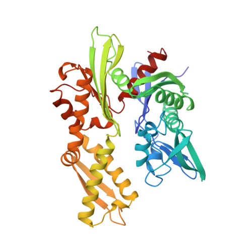
Three-dimensional structure of the ATPase fragment of a 70K heat-shock cognate protein.

Flaherty, K.M.DeLuca-Flaherty, C.McKay, D.B.

(1990) Nature 346: 623-628

  • DOI: https://doi.org/10.1038/346623a0
  • Primary Citation of Related Structures:  
    3HSC

  • PubMed Abstract: 
  • The three-dimensional structure of the amino-terminal 44K ATPase fragment of the 70K bovine heat-shock cognate protein has been solved to a resolution of 2.2 A. The ATPase fragment has two structural lobes with a deep cleft between them; ATP binds at the base of the cleft ...

    The three-dimensional structure of the amino-terminal 44K ATPase fragment of the 70K bovine heat-shock cognate protein has been solved to a resolution of 2.2 A. The ATPase fragment has two structural lobes with a deep cleft between them; ATP binds at the base of the cleft. Surprisingly, the nucleotide-binding 'core' of the ATPase fragment has a tertiary structure similar to that of hexokinase, although the remainder of the structures of the two proteins are completely dissimilar, suggesting that both the phosphotransferase mechanism and the substrate-induced conformational change intrinsic to the hexokinases may be used by the 70K heat shock-related proteins.


    Related Citations: 
    • How Potassium Affects the Activity of the Molecular Chaperone Hsc70 II. Potassium Binds Specifically in the ATPase Active Site
      Wilbanks, S.M., Mckay, D.B.
      (1995) J Biol Chem 270: 2251
    • How Potassium Affects the Activity of the Molecular Chaperone Hsc70 I. Potassium is Required for Optimal ATPase Activity
      O'Brien, M.C., Mckay, D.B.
      (1995) J Biol Chem 270: 2247
    • Structural Basis of the 70-Kilodalton Heat Shock Cognate Protein ATP Hydrolytic Activity, II. Structure of the Active Site with Adp or ATP Bound to Wild Type and Mutant ATPase Fragment
      Flaherty, K.M., Wilbanks, S.M., Deluca-Flaherty, C., Mckay, D.B.
      (1994) J Biol Chem 269: 12899

    Organizational Affiliation

    Beckman Laboratories for Structural Biology, Department of Cell Biology, Stanford University School of Medicine, California 94305-5400.


Macromolecules
Find similar proteins by:  (by identity cutoff)  |  3D Structure
Entity ID: 1
MoleculeChainsSequence LengthOrganismDetailsImage
HEAT-SHOCK COGNATE 7OKD PROTEIN386Bos taurusMutation(s): 0 
Gene Names: HSPA8HSC70
EC: 3.6.1.3 (PDB Primary Data), 3.6.4.10 (UniProt)
UniProt
Find proteins for P19120 (Bos taurus)
Explore P19120 
Go to UniProtKB:  P19120
Entity Groups  
Sequence Clusters30% Identity50% Identity70% Identity90% Identity95% Identity100% Identity
UniProt GroupP19120
Protein Feature View
Expand
  • Reference Sequence
Small Molecules
Ligands 4 Unique
IDChainsName / Formula / InChI Key2D Diagram3D Interactions
ADP
Query on ADP

Download Ideal Coordinates CCD File 
F [auth A]ADENOSINE-5'-DIPHOSPHATE
C10 H15 N5 O10 P2
XTWYTFMLZFPYCI-KQYNXXCUSA-N
 Ligand Interaction
PO4
Query on PO4

Download Ideal Coordinates CCD File 
C [auth A]PHOSPHATE ION
O4 P
NBIIXXVUZAFLBC-UHFFFAOYSA-K
 Ligand Interaction
MG
Query on MG

Download Ideal Coordinates CCD File 
B [auth A]MAGNESIUM ION
Mg
JLVVSXFLKOJNIY-UHFFFAOYSA-N
 Ligand Interaction
NA
Query on NA

Download Ideal Coordinates CCD File 
D [auth A],
E [auth A]
SODIUM ION
Na
FKNQFGJONOIPTF-UHFFFAOYSA-N
 Ligand Interaction
Binding Affinity Annotations 
IDSourceBinding Affinity
ADP BindingDB:  3HSC Kd: min: 320, max: 324 (nM) from 2 assay(s)
IC50: 280 (nM) from 1 assay(s)
Experimental Data & Validation

Experimental Data

  • Method: X-RAY DIFFRACTION
  • Resolution: 1.93 Å
  • R-Value Work: 0.210 
  • R-Value Observed: 0.210 
  • Space Group: P 21 21 21
Unit Cell:
Length ( Å )Angle ( ˚ )
a = 145.3α = 90
b = 65β = 90
c = 46.9γ = 90
Software Package:
Software NamePurpose
X-PLORmodel building
X-PLORrefinement
X-PLORphasing

Structure Validation

View Full Validation Report


Entry History 

Deposition Data

Revision History  (Full details and data files)

  • Version 1.0: 1995-07-10
    Type: Initial release
  • Version 1.1: 2008-03-25
    Changes: Version format compliance
  • Version 1.2: 2011-07-13
    Changes: Version format compliance