3HL5 | pdb_00003hl5

Crystal structure of XIAP BIR3 with CS3


Experimental Data Snapshot

  • Method: X-RAY DIFFRACTION
  • Resolution: 1.80 Å
  • R-Value Free: 
    0.209 (Depositor), 0.209 (DCC) 
  • R-Value Work: 
    0.176 (Depositor), 0.175 (DCC) 
  • R-Value Observed: 
    0.179 (Depositor) 

Starting Model: experimental
View more details

wwPDB Validation 3D Report Full Report

Validation slider image for 3HL5

Ligand Structure Quality Assessment 


This is version 1.2 of the entry. See complete history

Literature

Antagonism of c-IAP and XIAP proteins is required for efficient induction of cell death by small-molecule IAP antagonists.

Ndubaku, C.Varfolomeev, E.Wang, L.Zobel, K.Lau, K.Elliott, L.O.Maurer, B.Fedorova, A.V.Dynek, J.N.Koehler, M.Hymowitz, S.G.Tsui, V.Deshayes, K.Fairbrother, W.J.Flygare, J.A.Vucic, D.

(2009) ACS Chem Biol 4: 557-566

  • DOI: https://doi.org/10.1021/cb900083m
  • Primary Citation Related Structures: 
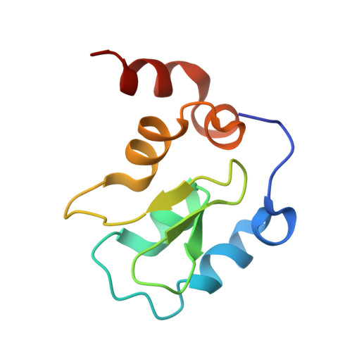
    3HL5

  • PubMed Abstract: 

    The inhibitor of apoptosis (IAP) proteins are critical regulators of cancer cell survival, which makes them attractive targets for therapeutic intervention in cancers. Herein, we describe the structure-based design of IAP antagonists with high affinities and selectivity (>2000-fold) for c-IAP1 over XIAP and their functional characterization as activators of apoptosis in tumor cells. Although capable of inducing cell death and preventing clonogenic survival, c-IAP-selective antagonists are significantly less potent in promoting apoptosis when compared to pan-selective compounds. However, both pan-IAP- and c-IAP-selective antagonists stimulate c-IAP1 and c-IAP2 degradation and activation of NF-kappaB pathways with comparable potencies. Therefore, although compounds that specifically target c-IAP1 and c-IAP2 are capable of inducing apoptosis, antagonism of the c-IAP proteins and XIAP is required for efficient induction of cancer cell death by IAP antagonists.


  • Organizational Affiliation
    • Departments of Medicinal Chemistry and Protein Engineering, Genentech, Inc., South San Francisco, CA 94080, USA.

Macromolecule Content 

  • Total Structure Weight: 23.1 kDa 
  • Atom Count: 1,728 
  • Modeled Residue Count: 184 
  • Deposited Residue Count: 190 
  • Unique protein chains: 1

Macromolecules

Find similar proteins by:|  3D Structure
Entity ID: 1
MoleculeChains  Sequence LengthOrganismDetailsImage
Baculoviral IAP repeat-containing protein 4
A, B
95Homo sapiensMutation(s): 0 
Gene Names: API3BIRC4IAP3XIAP
EC: 6.3.2 (PDB Primary Data), 2.3.2.27 (UniProt)
UniProt & NIH Common Fund Data Resources
Find proteins for P98170 (Homo sapiens)
Explore P98170 
Go to UniProtKB:  P98170
PHAROS:  P98170
GTEx:  ENSG00000101966 
Entity Groups
Sequence Clusters30% Identity50% Identity70% Identity90% Identity95% Identity100% Identity
UniProt GroupP98170
Sequence Annotations
Expand
Reference Sequence

Experimental Data & Validation

Experimental Data

  • Method: X-RAY DIFFRACTION
  • Resolution: 1.80 Å
  • R-Value Free:  0.209 (Depositor), 0.209 (DCC) 
  • R-Value Work:  0.176 (Depositor), 0.175 (DCC) 
  • R-Value Observed: 0.179 (Depositor) 
Space Group: C 1 2 1
Unit Cell:
Length ( Å )Angle ( ˚ )
a = 103.913α = 90
b = 39.94β = 100
c = 49.929γ = 90
Software Package:
Software NamePurpose
BOSdata collection
PHASERphasing
REFMACrefinement
HKL-2000data reduction
HKL-2000data scaling

Structure Validation

View Full Validation Report


Ligand Structure Quality Assessment 


Entry History 

Deposition Data

Revision History  (Full details and data files)

  • Version 1.0: 2009-06-16
    Type: Initial release
  • Version 1.1: 2011-07-13
    Changes: Advisory, Refinement description, Version format compliance
  • Version 1.2: 2023-09-06
    Changes: Data collection, Database references, Derived calculations, Refinement description