3HIG | pdb_00003hig

Crystal structure of human diamine oxidase in complex with the inhibitor berenil


Experimental Data Snapshot

  • Method: X-RAY DIFFRACTION
  • Resolution: 2.09 Å
  • R-Value Free: 
    0.210 (Depositor), 0.212 (DCC) 
  • R-Value Work: 
    0.168 (Depositor), 0.173 (DCC) 
  • R-Value Observed: 
    0.171 (Depositor) 

Starting Model: experimental
View more details

wwPDB Validation 3D Report Full Report

Validation slider image for 3HIG

Ligand Structure Quality Assessment 


This is version 2.2 of the entry. See complete history

Literature

Structure and inhibition of human diamine oxidase

McGrath, A.P.Hilmer, K.M.Collyer, C.A.Shepard, E.M.Elmore, B.O.Brown, D.E.Dooley, D.M.Guss, J.M.

(2009) Biochemistry 48: 9810-9822

  • DOI: https://doi.org/10.1021/bi9014192
  • Primary Citation Related Structures: 
    3HI7, 3HIG, 3HII

  • PubMed Abstract: 

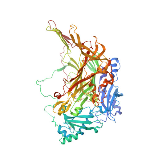
    Humans have three functioning genes that encode copper-containing amine oxidases. The product of the AOC1 gene is a so-called diamine oxidase (hDAO), named for its substrate preference for diamines, particularly histamine. hDAO has been cloned and expressed in insect cells and the structure of the native enzyme determined by X-ray crystallography to a resolution of 1.8 A. The homodimeric structure has the archetypal amine oxidase fold. Two active sites, one in each subunit, are characterized by the presence of a copper ion and a topaquinone residue formed by the post-translational modification of a tyrosine. Although hDAO shares 37.9% sequence identity with another human copper amine oxidase, semicarbazide sensitive amine oxidase or vascular adhesion protein-1, its substrate binding pocket and entry channel are distinctly different in accord with the different substrate specificities. The structures of two inhibitor complexes of hDAO, berenil and pentamidine, have been refined to resolutions of 2.1 and 2.2 A, respectively. They bind noncovalently in the active-site channel. The inhibitor binding suggests that an aspartic acid residue, conserved in all diamine oxidases but absent from other amine oxidases, is responsible for the diamine specificity by interacting with the second amino group of preferred diamine substrates.


  • Organizational Affiliation
    • School of Molecular and Microbial Biosciences, University of Sydney, Sydney, NSW 2006, Australia.

Macromolecule Content 

  • Total Structure Weight: 170.58 kDa 
  • Atom Count: 12,772 
  • Modeled Residue Count: 1,425 
  • Deposited Residue Count: 1,462 
  • Unique protein chains: 1

Macromolecules

Find similar proteins by:|  3D Structure
Entity ID: 1
MoleculeChains  Sequence LengthOrganismDetailsImage
Amiloride-sensitive amine oxidase
A, B
731Homo sapiensMutation(s): 1 
Gene Names: ABP1
EC: 1.4.3.22
UniProt & NIH Common Fund Data Resources
Find proteins for P19801 (Homo sapiens)
Explore P19801 
Go to UniProtKB:  P19801
PHAROS:  P19801
GTEx:  ENSG00000002726 
Entity Groups
Sequence Clusters30% Identity50% Identity70% Identity90% Identity95% Identity100% Identity
UniProt GroupP19801
Glycosylation
Glycosylation Sites: 3Go to GlyGen: P19801-1
Sequence Annotations
Expand
Reference Sequence

Oligosaccharides

Help  
Entity ID: 2
MoleculeChains Length2D Diagram GlycosylationD Interactions
beta-D-mannopyranose-(1-4)-2-acetamido-2-deoxy-beta-D-glucopyranose-(1-4)-2-acetamido-2-deoxy-beta-D-glucopyranose
C
3N-Glycosylation
Glycosylation Resources
GlyTouCan: G15407YE
GlyCosmos: G15407YE
GlyGen: G15407YE
Entity ID: 3
MoleculeChains Length2D Diagram GlycosylationD Interactions
2-acetamido-2-deoxy-beta-D-glucopyranose-(1-4)-2-acetamido-2-deoxy-beta-D-glucopyranose
D, E, F, G, H
2N-Glycosylation
Glycosylation Resources
GlyTouCan: G42666HT
GlyCosmos: G42666HT
GlyGen: G42666HT

Small Molecules

Ligands 4 Unique
IDChains Name / Formula / InChI Key2D Diagram3D Interactions
BRN

Query on BRN


Download:Ideal Coordinates CCD File
M [auth A],
Q [auth B]
BERENIL
C14 H15 N7
XNYZHCFCZNMTFY-UHFFFAOYSA-N
GOL

Query on GOL


Download:Ideal Coordinates CCD File
L [auth A]GLYCEROL
C3 H8 O3
PEDCQBHIVMGVHV-UHFFFAOYSA-N
CU

Query on CU


Download:Ideal Coordinates CCD File
I [auth A],
N [auth B]
COPPER (II) ION
Cu
JPVYNHNXODAKFH-UHFFFAOYSA-N
CA

Query on CA


Download:Ideal Coordinates CCD File
J [auth A],
K [auth A],
O [auth B],
P [auth B]
CALCIUM ION
Ca
BHPQYMZQTOCNFJ-UHFFFAOYSA-N
Modified Residues  1 Unique
IDChains TypeFormula2D DiagramParent
TPQ
Query on TPQ
A, B
L-PEPTIDE LINKINGC9 H9 N O5TYR
Binding Affinity Annotations 
IDSourceBinding Affinity
BRN BindingDB:  3HIG Ki: 13 (nM) from 1 assay(s)

Experimental Data & Validation

Experimental Data

  • Method: X-RAY DIFFRACTION
  • Resolution: 2.09 Å
  • R-Value Free:  0.210 (Depositor), 0.212 (DCC) 
  • R-Value Work:  0.168 (Depositor), 0.173 (DCC) 
  • R-Value Observed: 0.171 (Depositor) 
Space Group: P 21 21 21
Unit Cell:
Length ( Å )Angle ( ˚ )
a = 92.662α = 90
b = 94.884β = 90
c = 196.411γ = 90
Software Package:
Software NamePurpose
DENZOdata reduction
SCALEPACKdata scaling
REFMACrefinement
PDB_EXTRACTdata extraction
Blu-Icedata collection
HKL-2000data reduction
HKL-2000data scaling
REFMACphasing

Structure Validation

View Full Validation Report


Ligand Structure Quality Assessment 


Entry History 

Deposition Data

Revision History  (Full details and data files)

  • Version 1.0: 2009-10-20
    Type: Initial release
  • Version 1.1: 2011-07-13
    Changes: Derived calculations, Non-polymer description, Version format compliance
  • Version 1.2: 2017-11-01
    Changes: Refinement description
  • Version 2.0: 2020-07-29
    Type: Remediation
    Reason: Carbohydrate remediation
    Changes: Atomic model, Data collection, Derived calculations, Structure summary
  • Version 2.1: 2021-11-10
    Changes: Database references, Structure summary
  • Version 2.2: 2023-11-01
    Changes: Data collection, Refinement description