3HE3

Crystal Structure of UDP-galactopyranose mutase in complex with UDP


Experimental Data Snapshot

  • Method: X-RAY DIFFRACTION
  • Resolution: 2.40 Å
  • R-Value Free: 0.254 
  • R-Value Work: 0.208 
  • R-Value Observed: 0.211 

wwPDB Validation   3D Report Full Report


Ligand Structure Quality Assessment 


This is version 1.2 of the entry. See complete history


Literature

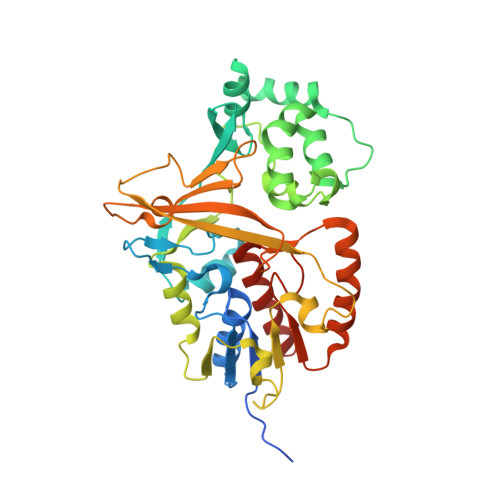
Structural basis of substrate binding to UDP-galactopyranose mutase: crystal structures in the reduced and oxidized state complexed with UDP-galactopyranose and UDP.

Partha, S.K.van Straaten, K.E.Sanders, D.A.

(2009) J Mol Biol 394: 864-877

  • DOI: https://doi.org/10.1016/j.jmb.2009.10.013
  • Primary Citation of Related Structures:  
    3HDQ, 3HDY, 3HE3

  • PubMed Abstract: 

    D-Galactofuranose (Galf) residues are found in the cell walls of pathogenic microbes such as Mycobacterium tuberculosis, and are essential for viability. UDP-galactopyranose mutase (UGM) is a unique flavo-enzyme that catalyzes the reversible conversion of UDP-galactopyranose (UDP-Galp) and UDP-galactofuranose (UDP-Galf). UDP-Galf is the active precursor of Galf residues found in cell walls. Despite the wealth of biochemical/mechanistic data generated for UGM, the structural basis of substrate binding is still lacking. Here, we report the crystal structures of UGM from Deinococcus radiodurans (drUGM) in complex with its natural substrate (UDP-Galp) and UDP. Crystal structures of drUGM:UDP-Galp complexes with oxidized and reduced FAD were determined at 2.36 A and 2.50 A resolution, respectively. The substrate is buried in the active site in an unusual folded conformation and the anomeric carbon of the galactose is at a favorable distance (2.8 A) from N5 of FAD to form an FAD-galactose adduct. The mobile loops in the substrate complex structure exist in a closed conformation. The drUGM-UDP complex structure was determined at 2.55 A resolution and its overall structure is identical with that of the oxidized and reduced complexes, including the conformation of the mobile loops. Comparison with the recently reported UGM:UDP-glucose complex structure reveals key differences and the structures reported here are likely to represent the productive/active conformation of UGM. These structures provide valuable insights into substrate recognition and a basis for understanding the mechanism. These complex structures may serve as a platform for structure-guided design of inhibitors of UGM.


  • Organizational Affiliation

    Department of Chemistry, 110 Science Place, University of Saskatchewan, Saskatoon, Canada S7N 5C9.


Macromolecules
Find similar proteins by:  (by identity cutoff)  |  3D Structure
Entity ID: 1
MoleculeChains Sequence LengthOrganismDetailsImage
UDP-galactopyranose mutase
A, B, C, D, E
A, B, C, D, E, F, G, H, I, J
397Deinococcus radioduransMutation(s): 0 
Gene Names: DR_A0367
EC: 5.4.99.9
UniProt
Find proteins for Q9RYF1 (Deinococcus radiodurans (strain ATCC 13939 / DSM 20539 / JCM 16871 / CCUG 27074 / LMG 4051 / NBRC 15346 / NCIMB 9279 / VKM B-1422 / R1))
Explore Q9RYF1 
Go to UniProtKB:  Q9RYF1
Entity Groups  
Sequence Clusters30% Identity50% Identity70% Identity90% Identity95% Identity100% Identity
UniProt GroupQ9RYF1
Sequence Annotations
Expand
  • Reference Sequence
Small Molecules
Ligands 2 Unique
IDChains Name / Formula / InChI Key2D Diagram3D Interactions
FAD
Query on FAD

Download Ideal Coordinates CCD File 
BA [auth I]
DA [auth J]
L [auth A]
N [auth B]
P [auth C]
BA [auth I],
DA [auth J],
L [auth A],
N [auth B],
P [auth C],
R [auth D],
T [auth E],
V [auth F],
X [auth G],
Z [auth H]
FLAVIN-ADENINE DINUCLEOTIDE
C27 H33 N9 O15 P2
VWWQXMAJTJZDQX-UYBVJOGSSA-N
UDP
Query on UDP

Download Ideal Coordinates CCD File 
AA [auth I]
CA [auth J]
K [auth A]
M [auth B]
O [auth C]
AA [auth I],
CA [auth J],
K [auth A],
M [auth B],
O [auth C],
Q [auth D],
S [auth E],
U [auth F],
W [auth G],
Y [auth H]
URIDINE-5'-DIPHOSPHATE
C9 H14 N2 O12 P2
XCCTYIAWTASOJW-XVFCMESISA-N
Experimental Data & Validation

Experimental Data

  • Method: X-RAY DIFFRACTION
  • Resolution: 2.40 Å
  • R-Value Free: 0.254 
  • R-Value Work: 0.208 
  • R-Value Observed: 0.211 
  • Space Group: P 21 21 21
Unit Cell:
Length ( Å )Angle ( ˚ )
a = 134.05α = 90
b = 176.874β = 90
c = 222.92γ = 90
Software Package:
Software NamePurpose
PHENIXrefinement

Structure Validation

View Full Validation Report


Ligand Structure Quality Assessment 


Entry History 

Deposition Data

Revision History  (Full details and data files)

  • Version 1.0: 2009-12-29
    Type: Initial release
  • Version 1.1: 2011-07-13
    Changes: Version format compliance
  • Version 1.2: 2023-09-06
    Changes: Data collection, Database references, Derived calculations, Refinement description