3H8H | pdb_00003h8h

Structure of the C-terminal domain of human RNF2/RING1B;


Experimental Data Snapshot

  • Method: X-RAY DIFFRACTION
  • Resolution: 2.00 Å
  • R-Value Free: 
    0.244 (Depositor), 0.274 (DCC) 
  • R-Value Work: 
    0.200 (Depositor), 0.224 (DCC) 
  • R-Value Observed: 
    0.202 (Depositor) 

wwPDB Validation 3D Report Full Report

Validation slider image for 3H8H

This is version 1.3 of the entry. See complete history

Literature

Ring1B contains a ubiquitin-like docking module for interaction with Cbx proteins.

Bezsonova, I.Walker, J.R.Bacik, J.P.Duan, S.Dhe-Paganon, S.Arrowsmith, C.H.

(2009) Biochemistry 48: 10542-10548

  • DOI: https://doi.org/10.1021/bi901131u
  • Primary Citation Related Structures: 
    3H8H

  • PubMed Abstract: 

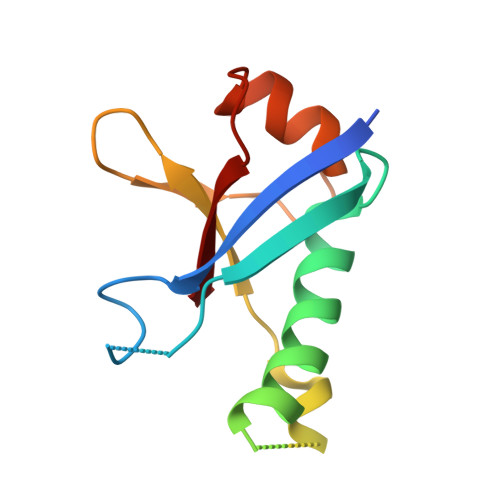
    Polycomb group (PcG) proteins are a special set of repressive transcription factors involved in epigenetic modifications of chromatin. They form two functionally distinct groups of catalytically active complexes: Polycomb repressive complex 1 (PRC1) and 2 (PRC2). The PRC1 complex is an important yet poorly characterized multiprotein histone ubiquitylation machine responsible for maintaining transcriptionally silent states of genes through histone H2A K119 modification. The Ring domain containing subunits of PRC1 also have substrate-targeting domains that interact with Cbx proteins, which have been implicated in chromatin and RNA binding. In this work, we present a high resolution structure of the C-terminal domain of Ring1B, revealing a variant ubiquitin-like fold with a distinct conserved surface region. On the basis of crystal structure and mutational analysis of this domain we show that the conserved surface is responsible for interaction with Cbx members of the PRC1 and homodimer formation. These data suggest a mechanism by which Ring1B serves as an adaptor that mediates binding between the members of the PRC1 complex and the nucleosome.


  • Organizational Affiliation
    • Structural Genomics Consortium, University of Toronto, 101 College Street, Toronto, Ontario, M5G 1L5, Canada.

Macromolecule Content 

  • Total Structure Weight: 13.28 kDa 
  • Atom Count: 833 
  • Modeled Residue Count: 92 
  • Deposited Residue Count: 112 
  • Unique protein chains: 1

Macromolecules

Find similar proteins by:|  3D Structure
Entity ID: 1
MoleculeChains  Sequence LengthOrganismDetailsImage
E3 ubiquitin-protein ligase RING2112Homo sapiensMutation(s): 0 
Gene Names: BAP1DINGHIPI3RING1BRNF2
EC: 6.3.2 (PDB Primary Data), 2.3.2.27 (UniProt)
UniProt & NIH Common Fund Data Resources
Find proteins for Q99496 (Homo sapiens)
Explore Q99496 
Go to UniProtKB:  Q99496
PHAROS:  Q99496
GTEx:  ENSG00000121481 
Entity Groups
Sequence Clusters30% Identity50% Identity70% Identity90% Identity95% Identity100% Identity
UniProt GroupQ99496
Sequence Annotations
Expand
Reference Sequence

Experimental Data & Validation

Experimental Data

  • Method: X-RAY DIFFRACTION
  • Resolution: 2.00 Å
  • R-Value Free:  0.244 (Depositor), 0.274 (DCC) 
  • R-Value Work:  0.200 (Depositor), 0.224 (DCC) 
  • R-Value Observed: 0.202 (Depositor) 
Space Group: P 65 2 2
Unit Cell:
Length ( Å )Angle ( ˚ )
a = 72.169α = 90
b = 72.169β = 90
c = 90.966γ = 120
Software Package:
Software NamePurpose
SBC-Collectdata collection
SOLVEphasing
RESOLVEmodel building
REFMACrefinement
HKL-2000data reduction
HKL-2000data scaling
RESOLVEphasing

Structure Validation

View Full Validation Report


Entry History 

Revision History  (Full details and data files)

  • Version 1.0: 2009-06-23
    Type: Initial release
  • Version 1.1: 2011-07-13
    Changes: Non-polymer description, Version format compliance
  • Version 1.2: 2017-11-01
    Changes: Refinement description
  • Version 1.3: 2024-02-21
    Changes: Data collection, Database references, Derived calculations