3GZU

VP7 recoated rotavirus DLP


Experimental Data Snapshot

  • Method: ELECTRON MICROSCOPY
  • Resolution: 3.80 Å
  • Aggregation State: PARTICLE 
  • Reconstruction Method: SINGLE PARTICLE 

wwPDB Validation   3D Report Full Report


This is version 1.4 of the entry. See complete history


Literature

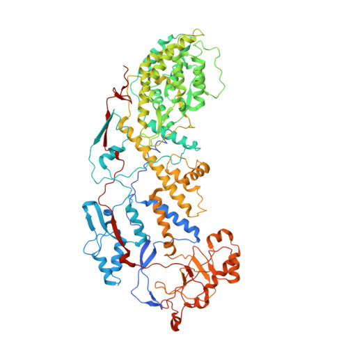
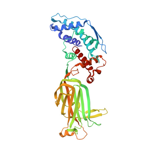
Molecular interactions in rotavirus assembly and uncoating seen by high-resolution cryo-EM.

Chen, J.Z.Settembre, E.C.Aoki, S.T.Zhang, X.Bellamy, A.R.Dormitzer, P.R.Harrison, S.C.Grigorieff, N.

(2009) Proc Natl Acad Sci U S A 106: 10644-10648

  • DOI: https://doi.org/10.1073/pnas.0904024106
  • Primary Citation of Related Structures:  
    3GZT, 3GZU

  • PubMed Abstract: 

    Rotaviruses, major causes of childhood gastroenteritis, are nonenveloped, icosahedral particles with double-strand RNA genomes. By the use of electron cryomicroscopy and single-particle reconstruction, we have visualized a rotavirus particle comprising the inner capsid coated with the trimeric outer-layer protein, VP7, at a resolution (4 A) comparable with that of X-ray crystallography. We have traced the VP7 polypeptide chain, including parts not seen in its X-ray crystal structure. The 3 well-ordered, 30-residue, N-terminal "arms" of each VP7 trimer grip the underlying trimer of VP6, an inner-capsid protein. Structural differences between free and particle-bound VP7 and between free and VP7-coated inner capsids may regulate mRNA transcription and release. The Ca(2+)-stabilized VP7 intratrimer contact region, which presents important neutralizing epitopes, is unaltered upon capsid binding.


  • Organizational Affiliation

    Rosenstiel Basic Medical Research Center, Brandeis University, Waltham, MA 02454, USA.


Macromolecules
Find similar proteins by:  (by identity cutoff)  |  3D Structure
Entity ID: 1
MoleculeChains Sequence LengthOrganismDetailsImage
Inner capsid protein VP2
A, B
800Rotavirus AMutation(s): 0 
UniProt
Find proteins for B2BMD1 (Rotavirus A)
Explore B2BMD1 
Go to UniProtKB:  B2BMD1
Entity Groups  
Sequence Clusters30% Identity50% Identity70% Identity90% Identity95% Identity100% Identity
UniProt GroupB2BMD1
Sequence Annotations
Expand
  • Reference Sequence
Find similar proteins by:  (by identity cutoff)  |  3D Structure
Entity ID: 2
MoleculeChains Sequence LengthOrganismDetailsImage
Intermediate capsid protein VP6
C, D, E, F, G
C, D, E, F, G, H, I, J, K, L, M, N, O
397Simian rotavirus A strain RRVMutation(s): 0 
UniProt
Find proteins for P04509 (Rotavirus A (strain RVA/Cow/France/RF/1975/G6P6[1]))
Explore P04509 
Go to UniProtKB:  P04509
Entity Groups  
Sequence Clusters30% Identity50% Identity70% Identity90% Identity95% Identity100% Identity
UniProt GroupP04509
Sequence Annotations
Expand
  • Reference Sequence
Experimental Data & Validation

Experimental Data

  • Method: ELECTRON MICROSCOPY
  • Resolution: 3.80 Å
  • Aggregation State: PARTICLE 
  • Reconstruction Method: SINGLE PARTICLE 

Structure Validation

View Full Validation Report


Entry History 

Deposition Data

Revision History  (Full details and data files)

  • Version 1.0: 2009-07-14
    Type: Initial release
  • Version 1.1: 2011-07-13
    Changes: Version format compliance
  • Version 1.2: 2019-11-06
    Changes: Data collection, Other
  • Version 1.3: 2019-11-13
    Changes: Data collection, Other
  • Version 1.4: 2024-02-21
    Changes: Data collection, Database references, Derived calculations