3GM2

Crystal Structure of the Focal Adhesion Targeting (FAT) Domain of Pyk2


Experimental Data Snapshot

  • Method: X-RAY DIFFRACTION
  • Resolution: 2.71 Å
  • R-Value Free: 0.295 
  • R-Value Work: 0.261 
  • R-Value Observed: 0.261 

wwPDB Validation   3D Report Full Report


This is version 1.2 of the entry. See complete history


Literature

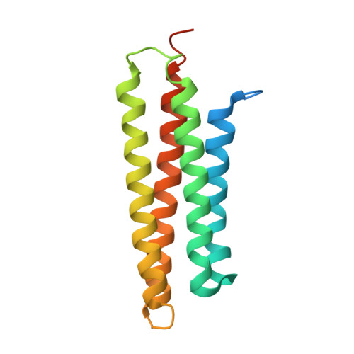
Crystal structures of free and ligand-bound focal adhesion targeting domain of Pyk2

Lulo, J.Yuzawa, S.Schlessinger, J.

(2009) Biochem Biophys Res Commun 383: 347-352

  • DOI: https://doi.org/10.1016/j.bbrc.2009.04.011
  • Primary Citation of Related Structures:  
    3GM1, 3GM2, 3GM3

  • PubMed Abstract: 

    Focal adhesion targeting (FAT) domains target the non-receptor tyrosine kinases FAK and Pyk2 to cellular focal adhesion areas, where the signaling molecule paxillin is also located. Here, we report the crystal structures of the Pyk2 FAT domain alone or in complex with paxillin LD4 peptides. The overall structure of Pyk2-FAT is an antiparallel four-helix bundle with an up-down, up-down, right-handed topology. In the LD4-bound FAT complex, two paxillin LD4 peptides interact with two opposite sides of Pyk2-FAT, at the surfaces of the alpha1alpha4 and alpha2alpha3 helices of each FAT molecule. We also demonstrate that, while paxillin is phosphorylated by Pyk2, complex formation between Pyk2 and paxillin does not depend on Pyk2 tyrosine kinase activity. These experiments reveal the structural basis underlying the selectivity of paxillin LD4 binding to the Pyk2 FAT domain and provide insights about the molecular details which influence the different behavior of these two closely-related kinases.


  • Organizational Affiliation

    Department of Pharmacology, Yale University School of Medicine, New Haven, CT 06520, USA.


Macromolecules
Find similar proteins by:  (by identity cutoff)  |  3D Structure
Entity ID: 1
MoleculeChains Sequence LengthOrganismDetailsImage
Protein tyrosine kinase 2 beta153Homo sapiensMutation(s): 1 
EC: 2.7.10.2
UniProt & NIH Common Fund Data Resources
Find proteins for Q14289 (Homo sapiens)
Explore Q14289 
Go to UniProtKB:  Q14289
PHAROS:  Q14289
GTEx:  ENSG00000120899 
Entity Groups  
Sequence Clusters30% Identity50% Identity70% Identity90% Identity95% Identity100% Identity
UniProt GroupQ14289
Sequence Annotations
Expand
  • Reference Sequence
Experimental Data & Validation

Experimental Data

  • Method: X-RAY DIFFRACTION
  • Resolution: 2.71 Å
  • R-Value Free: 0.295 
  • R-Value Work: 0.261 
  • R-Value Observed: 0.261 
  • Space Group: I 41 2 2
Unit Cell:
Length ( Å )Angle ( ˚ )
a = 82.547α = 90
b = 82.547β = 90
c = 139.528γ = 90
Software Package:
Software NamePurpose
CBASSdata collection
CNSrefinement
HKL-2000data reduction
HKL-2000data scaling
CNSphasing

Structure Validation

View Full Validation Report


Entry History 

Deposition Data

Revision History  (Full details and data files)

  • Version 1.0: 2009-05-05
    Type: Initial release
  • Version 1.1: 2011-07-13
    Changes: Version format compliance
  • Version 1.2: 2021-11-10
    Changes: Database references