3GHG | pdb_00003ghg

Crystal Structure of Human Fibrinogen


Experimental Data Snapshot

  • Method: X-RAY DIFFRACTION
  • Resolution: 2.90 Å
  • R-Value Free: 
    0.309 (Depositor), 0.311 (DCC) 
  • R-Value Work: 
    0.252 (Depositor), 0.259 (DCC) 
  • R-Value Observed: 
    0.255 (Depositor) 

wwPDB Validation 3D Report Full Report

Validation slider image for 3GHG

This is version 3.1 of the entry. See complete history

Literature

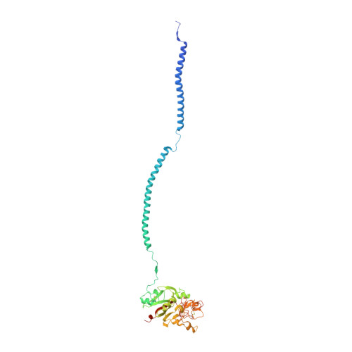
Crystal structure of human fibrinogen.

Kollman, J.M.Pandi, L.Sawaya, M.R.Riley, M.Doolittle, R.F.

(2009) Biochemistry 48: 3877-3886

  • DOI: https://doi.org/10.1021/bi802205g
  • Primary Citation Related Structures: 
    3GHG

  • PubMed Abstract: 

    A crystal structure of human fibrinogen has been determined at approximately 3.3 A resolution. The protein was purified from human blood plasma, first by a cold ethanol precipitation procedure and then by stepwise chromatography on DEAE-cellulose. A product was obtained that was homogeneous on SDS-polyacrylamide gels. Nonetheless, when individual crystals used for X-ray diffraction were examined by SDS gel electrophoresis after data collection, two species of alpha chain were present, indicating that some proteolysis had occurred during the course of operations. Amino-terminal sequencing on post-X-ray crystals showed mostly intact native alpha- and gamma-chain sequences (the native beta chain is blocked). The overall structure differs from that of a native fibrinogen from chicken blood and those reported for a partially proteolyzed bovine fibrinogen in the nature of twist in the coiled-coil regions, likely due to weak forces imparted by unique crystal packing. As such, the structure adds to the inventory of possible conformations that may occur in solution. Other features include a novel interface with an antiparallel arrangement of beta chains and a unique tangential association of coiled coils from neighboring molecules. The carbohydrate groups attached to beta chains are unusually prominent, the full sweep of 11 sugar residues being positioned. As was the case for native chicken fibrinogen, no resolvable electron density could be associated with alphaC domains.


  • Organizational Affiliation
    • Department of Chemistry and Biochemistry and Division of Biology, University of California at San Diego, La Jolla, California 92093-0314, USA.

Macromolecule Content 

  • Total Structure Weight: 651.39 kDa 
  • Atom Count: 31,833 
  • Modeled Residue Count: 3,892 
  • Deposited Residue Count: 5,768 
  • Unique protein chains: 5

Macromolecules

Find similar proteins by:|  3D Structure
Entity ID: 1
MoleculeChains  Sequence LengthOrganismDetailsImage
Fibrinogen alpha chain
A, D, G, J
562Homo sapiensMutation(s): 0 
UniProt & NIH Common Fund Data Resources
Find proteins for P02671 (Homo sapiens)
Explore P02671 
Go to UniProtKB:  P02671
PHAROS:  P02671
GTEx:  ENSG00000171560 
Entity Groups
Sequence Clusters30% Identity50% Identity70% Identity90% Identity95% Identity100% Identity
UniProt GroupP02671
Sequence Annotations
Expand
Reference Sequence
Find similar proteins by:|  3D Structure
Entity ID: 2
MoleculeChains  Sequence LengthOrganismDetailsImage
Fibrinogen beta chain
B, E, H, K
461Homo sapiensMutation(s): 0 
UniProt & NIH Common Fund Data Resources
Find proteins for P02675 (Homo sapiens)
Explore P02675 
Go to UniProtKB:  P02675
PHAROS:  P02675
GTEx:  ENSG00000171564 
Entity Groups
Sequence Clusters30% Identity50% Identity70% Identity90% Identity95% Identity100% Identity
UniProt GroupP02675
Glycosylation
Glycosylation Sites: 1Go to GlyGen: P02675-1
Sequence Annotations
Expand
Reference Sequence
Find similar proteins by:|  3D Structure
Entity ID: 3
MoleculeChains  Sequence LengthOrganismDetailsImage
Fibrinogen gamma chain
C, F, I, L
411Homo sapiensMutation(s): 0 
UniProt & NIH Common Fund Data Resources
Find proteins for P02679 (Homo sapiens)
Explore P02679 
Go to UniProtKB:  P02679
PHAROS:  P02679
GTEx:  ENSG00000171557 
Entity Groups
Sequence Clusters30% Identity50% Identity70% Identity90% Identity95% Identity100% Identity
UniProt GroupP02679
Sequence Annotations
Expand
Reference Sequence
Find similar proteins by:  Sequence   |   3D Structure  
Entity ID: 4
MoleculeChains  Sequence LengthOrganismDetailsImage
A knob
M, N, Q, R
4N/AMutation(s): 0 
Find similar proteins by:  Sequence   |   3D Structure  
Entity ID: 5
MoleculeChains  Sequence LengthOrganismDetailsImage
B knob
O, P, S, T
4N/AMutation(s): 0 

Oligosaccharides

Help  
Entity ID: 6
MoleculeChains Length2D Diagram GlycosylationD Interactions
N-acetyl-alpha-neuraminic acid-(2-6)-beta-D-galactopyranose-(1-4)-2-acetamido-2-deoxy-beta-D-glucopyranose-(1-2)-alpha-D-mannopyranose-(1-3)-[N-acetyl-alpha-neuraminic acid-(2-6)-beta-D-galactopyranose-(1-4)-2-acetamido-2-deoxy-beta-D-glucopyranose-(1-2)-alpha-D-mannopyranose-(1-6)]beta-D-mannopyranose-(1-4)-2-acetamido-2-deoxy-beta-D-glucopyranose-(1-4)-2-acetamido-2-deoxy-beta-D-glucopyranose
U, V, X
11N-Glycosylation
Glycosylation Resources
GlyTouCan: G84467IZ
GlyCosmos: G84467IZ
GlyGen: G84467IZ
Entity ID: 7
MoleculeChains Length2D Diagram GlycosylationD Interactions
alpha-D-mannopyranose-(1-3)-[alpha-D-mannopyranose-(1-6)]beta-D-mannopyranose-(1-4)-2-acetamido-2-deoxy-beta-D-glucopyranose-(1-4)-2-acetamido-2-deoxy-beta-D-glucopyranose
W
5N-Glycosylation
Glycosylation Resources
GlyTouCan: G22768VO
GlyCosmos: G22768VO
GlyGen: G22768VO
Entity ID: 8
MoleculeChains Length2D Diagram GlycosylationD Interactions
2-acetamido-2-deoxy-beta-D-glucopyranose-(1-4)-2-acetamido-2-deoxy-beta-D-glucopyranose
Y
2N/A
Glycosylation Resources
GlyTouCan: G42666HT
GlyCosmos: G42666HT
GlyGen: G42666HT

Experimental Data & Validation

Experimental Data

  • Method: X-RAY DIFFRACTION
  • Resolution: 2.90 Å
  • R-Value Free:  0.309 (Depositor), 0.311 (DCC) 
  • R-Value Work:  0.252 (Depositor), 0.259 (DCC) 
  • R-Value Observed: 0.255 (Depositor) 
Space Group: P 1 21 1
Unit Cell:
Length ( Å )Angle ( ˚ )
a = 135.238α = 90
b = 94.866β = 94.81
c = 300.812γ = 90
Software Package:
Software NamePurpose
REFMACrefinement
PDB_EXTRACTdata extraction
ADSCdata collection
DENZOdata reduction
SCALEPACKdata scaling
PHASESphasing

Structure Validation

View Full Validation Report


Entry History 

Deposition Data

Revision History  (Full details and data files)

  • Version 1.0: 2009-05-19
    Type: Initial release
  • Version 1.1: 2011-07-13
    Changes: Advisory, Refinement description, Version format compliance
  • Version 2.0: 2020-07-29
    Type: Remediation
    Reason: Carbohydrate remediation
    Changes: Advisory, Atomic model, Data collection, Derived calculations, Structure summary
  • Version 3.0: 2023-05-03
    Changes: Advisory, Atomic model, Data collection, Database references, Derived calculations, Non-polymer description, Refinement description, Source and taxonomy, Structure summary
  • Version 3.1: 2024-11-06
    Changes: Data collection, Structure summary