3G5W | pdb_00003g5w

Crystal structure of Blue Copper Oxidase from Nitrosomonas europaea


Experimental Data Snapshot

  • Method: X-RAY DIFFRACTION
  • Resolution: 1.90 Å
  • R-Value Free: 
    0.197 (Depositor), 0.209 (DCC) 
  • R-Value Work: 
    0.166 (Depositor) 
  • R-Value Observed: 
    0.168 (Depositor) 

wwPDB Validation 3D Report Full Report

Validation slider image for 3G5W

This is version 1.2 of the entry. See complete history

Literature

Crystal structure of a two-domain multicopper oxidase: implications for the evolution of multicopper blue proteins.

Lawton, T.J.Sayavedra-Soto, L.A.Arp, D.J.Rosenzweig, A.C.

(2009) J Biological Chem 284: 10174-10180

  • DOI: https://doi.org/10.1074/jbc.M900179200
  • Primary Citation Related Structures: 
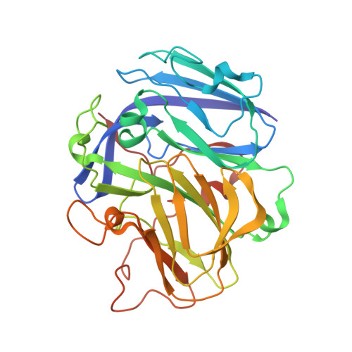
    3G5W

  • PubMed Abstract: 

    The two-domain multicopper oxidases are proposed to be key intermediates in the evolution of three-domain multicopper oxidases. A number of two-domain multicopper oxidases have been identified from genome sequences and are classified as type A, type B, or type C on the basis of the predicted location of the type 1 copper center. The crystal structure of blue copper oxidase, a type C two-domain multicopper oxidase from Nitrosomonas europaea, has been determined to 1.9 A resolution. Blue copper oxidase is a trimer, of which each subunit comprises two cupredoxin domains. Each subunit houses a type 1 copper site in domain 1 and a type 2/type 3 trinuclear copper cluster at the subunit-subunit interface. The coordination geometry at the trinuclear copper site is consistent with reduction of the copper ions. Although the overall architecture of blue copper oxidase is similar to nitrite reductases, detailed structural alignments show that the fold and domain orientation more closely resemble the three-domain multicopper oxidases. These observations have important implications for the evolution of nitrite reductases and multicopper oxidases.


  • Organizational Affiliation
    • Departments of Biochemistry, Molecular Biology, and Cell Biology and of Chemistry, Northwestern University, Evanston, Illinois 60208, USA.

Macromolecule Content 

  • Total Structure Weight: 218.81 kDa 
  • Atom Count: 16,620 
  • Modeled Residue Count: 1,908 
  • Deposited Residue Count: 1,908 
  • Unique protein chains: 1

Macromolecules

Find similar proteins by:|  3D Structure
Entity ID: 1
MoleculeChains  Sequence LengthOrganismDetailsImage
Multicopper oxidase type 1
A, B, C, D, E
A, B, C, D, E, F
318Nitrosomonas europaeaMutation(s): 0 
UniProt
Find proteins for Q82VX3 (Nitrosomonas europaea (strain ATCC 19718 / CIP 103999 / KCTC 2705 / NBRC 14298))
Explore Q82VX3 
Go to UniProtKB:  Q82VX3
Entity Groups
Sequence Clusters30% Identity50% Identity70% Identity90% Identity95% Identity100% Identity
UniProt GroupQ82VX3
Sequence Annotations
Expand
Reference Sequence

Small Molecules

Ligands 4 Unique
IDChains Name / Formula / InChI Key2D Diagram3D Interactions
C2O

Query on C2O


Download:Ideal Coordinates CCD File
AA [auth F]
K [auth B]
N [auth C]
P [auth C]
U [auth E]
AA [auth F],
K [auth B],
N [auth C],
P [auth C],
U [auth E],
Y [auth F]
CU-O-CU LINKAGE
Cu2 O
BERDEBHAJNAUOM-UHFFFAOYSA-N
GOL

Query on GOL


Download:Ideal Coordinates CCD File
I [auth A],
J [auth A],
T [auth D],
X [auth E]
GLYCEROL
C3 H8 O3
PEDCQBHIVMGVHV-UHFFFAOYSA-N
C1O

Query on C1O


Download:Ideal Coordinates CCD File
H [auth A]
L [auth B]
O [auth C]
S [auth D]
W [auth E]
H [auth A],
L [auth B],
O [auth C],
S [auth D],
W [auth E],
Z [auth F]
CU-O LINKAGE
Cu O
QPLDLSVMHZLSFG-UHFFFAOYSA-N
CU

Query on CU


Download:Ideal Coordinates CCD File
BA [auth F]
G [auth A]
M [auth B]
Q [auth C]
R [auth D]
BA [auth F],
G [auth A],
M [auth B],
Q [auth C],
R [auth D],
V [auth E]
COPPER (II) ION
Cu
JPVYNHNXODAKFH-UHFFFAOYSA-N

Experimental Data & Validation

Experimental Data

  • Method: X-RAY DIFFRACTION
  • Resolution: 1.90 Å
  • R-Value Free:  0.197 (Depositor), 0.209 (DCC) 
  • R-Value Work:  0.166 (Depositor) 
  • R-Value Observed: 0.168 (Depositor) 
Space Group: P 1
Unit Cell:
Length ( Å )Angle ( ˚ )
a = 76.14α = 81.48
b = 76.254β = 73.38
c = 105.145γ = 61.84
Software Package:
Software NamePurpose
REFMACrefinement
PDB_EXTRACTdata extraction

Structure Validation

View Full Validation Report


Entry History 

Deposition Data

Revision History  (Full details and data files)

  • Version 1.0: 2009-02-17
    Type: Initial release
  • Version 1.1: 2011-07-13
    Changes: Non-polymer description, Version format compliance
  • Version 1.2: 2024-02-21
    Changes: Data collection, Database references, Derived calculations