3FYI | pdb_00003fyi

Catalytic core subunits (I and II) of cytochrome C oxidase from Rhodobacter sphaeroides in the reduced state bound with cyanide


Experimental Data Snapshot

  • Method: X-RAY DIFFRACTION
  • Resolution: 2.20 Å
  • R-Value Free: 
    0.219 (Depositor), 0.216 (DCC) 
  • R-Value Work: 
    0.194 (Depositor), 0.193 (DCC) 
  • R-Value Observed: 
    0.195 (Depositor) 

Starting Model: experimental
View more details

wwPDB Validation 3D Report Full Report

Validation slider image for 3FYI

Ligand Structure Quality Assessment 


This is version 1.4 of the entry. See complete history

Literature

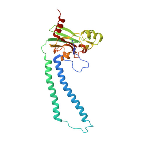
Redox dependent conformational changes in cytochrome c oxidase suggest a gating mechanism for proton uptake.

Qin, L.Liu, J.Mills, D.Proshlyakov, D.A.Hiser, C.Ferguson-Miller, S.

(2009) Biochemistry 48: 5121-5130

  • DOI: https://doi.org/10.1021/bi9001387
  • Primary Citation Related Structures: 
    3FYE, 3FYI

  • PubMed Abstract: 

    A role for conformational change in the coupling mechanism of cytochrome c oxidase is the subject of controversy. Relatively small conformational changes have been reported in comparisons of reduced and oxidized crystal structures of bovine oxidase but none in bacterial oxidases. Comparing the X-ray crystal structures of the reduced (at 2.15 A resolution) and oxidized forms of cytochrome c oxidase from Rhodobacter sphaeroides, we observe a displacement of heme a(3) involving both the porphyrin ring and the hydroxyl farnesyl tail, accompanied by protein movements in nearby regions, including the mid part of helix VIII of subunit I which harbors key residues of the K proton uptake path, K362 and T359. The conformational changes in the reduced form are reversible upon reoxidation. They result in an opening of the top of the K pathway and more ordered waters being resolved in that region, suggesting an access path for protons into the active site. In all high-resolution structures of oxidized R. sphaeroides cytochrome c oxidase, a water molecule is observed in the hydrophobic region above the top of the D path, strategically positioned to facilitate the connection of residue E286 of subunit I to the active site or to the proton pumping exit path. In the reduced and reduced plus cyanide structures, this water molecule disappears, implying disruption of proton conduction from the D path under conditions when the K path is open, thus providing a mechanism for alternating access to the active site.


  • Organizational Affiliation
    • Biochemistry and Molecular Biology Department, Michigan State University, East Lansing, Michigan 48824, USA.

Macromolecule Content 

  • Total Structure Weight: 196.73 kDa 
  • Atom Count: 13,601 
  • Modeled Residue Count: 1,578 
  • Deposited Residue Count: 1,656 
  • Unique protein chains: 2

Macromolecules

Find similar proteins by:|  3D Structure
Entity ID: 1
MoleculeChains  Sequence LengthOrganismDetailsImage
Cytochrome c oxidase subunit 1
A, C
566Cereibacter sphaeroidesMutation(s): 0 
Gene Names: ctaD
EC: 1.9.3.1 (PDB Primary Data), 7.1.1.9 (UniProt)
Membrane Entity: Yes 
UniProt
Find proteins for P33517 (Cereibacter sphaeroides)
Explore P33517 
Go to UniProtKB:  P33517
Entity Groups
Sequence Clusters30% Identity50% Identity70% Identity90% Identity95% Identity100% Identity
UniProt GroupP33517
Sequence Annotations
Expand
Reference Sequence
Find similar proteins by:|  3D Structure
Entity ID: 2
MoleculeChains  Sequence LengthOrganismDetailsImage
Cytochrome c oxidase subunit 2
B, D
262Cereibacter sphaeroidesMutation(s): 0 
Gene Names: ctaCcoxIIctaB
EC: 1.9.3.1 (PDB Primary Data), 7.1.1.9 (UniProt)
Membrane Entity: Yes 
UniProt
Find proteins for Q03736 (Cereibacter sphaeroides)
Explore Q03736 
Go to UniProtKB:  Q03736
Entity Groups
Sequence Clusters30% Identity50% Identity70% Identity90% Identity95% Identity100% Identity
UniProt GroupQ03736
Sequence Annotations
Expand
Reference Sequence

Small Molecules

Ligands 9 Unique
IDChains Name / Formula / InChI Key2D Diagram3D Interactions
HEA

Query on HEA


Download:Ideal Coordinates CCD File
BA [auth C],
CA [auth C],
E [auth A],
F [auth A]
HEME-A
C49 H56 Fe N4 O6
ZGGYGTCPXNDTRV-PRYGPKJJSA-L
DMU

Query on DMU


Download:Ideal Coordinates CCD File
AA [auth B]
IA [auth C]
JA [auth C]
K [auth A]
L [auth A]
AA [auth B],
IA [auth C],
JA [auth C],
K [auth A],
L [auth A],
M [auth A],
N [auth A],
RA [auth D],
UA [auth D],
Z [auth B]
DECYL-BETA-D-MALTOPYRANOSIDE
C22 H42 O11
WOQQAWHSKSSAGF-WXFJLFHKSA-N
TRD

Query on TRD


Download:Ideal Coordinates CCD File
HA [auth C]
KA [auth C]
LA [auth C]
MA [auth C]
O [auth A]
HA [auth C],
KA [auth C],
LA [auth C],
MA [auth C],
O [auth A],
P [auth A],
Q [auth A],
R [auth A],
S [auth A],
SA [auth D],
T [auth A],
TA [auth D]
TRIDECANE
C13 H28
IIYFAKIEWZDVMP-UHFFFAOYSA-N
HTO

Query on HTO


Download:Ideal Coordinates CCD File
U [auth B]HEPTANE-1,2,3-TRIOL
C7 H16 O3
HXYCHJFUBNTKQR-RNFRBKRXSA-N
CD

Query on CD


Download:Ideal Coordinates CCD File
PA [auth D],
QA [auth D],
X [auth B],
Y [auth B]
CADMIUM ION
Cd
WLZRMCYVCSSEQC-UHFFFAOYSA-N
CU1

Query on CU1


Download:Ideal Coordinates CCD File
DA [auth C]
G [auth A]
NA [auth D]
OA [auth D]
V [auth B]
DA [auth C],
G [auth A],
NA [auth D],
OA [auth D],
V [auth B],
W [auth B]
COPPER (I) ION
Cu
VMQMZMRVKUZKQL-UHFFFAOYSA-N
CA

Query on CA


Download:Ideal Coordinates CCD File
FA [auth C],
I [auth A]
CALCIUM ION
Ca
BHPQYMZQTOCNFJ-UHFFFAOYSA-N
CYN

Query on CYN


Download:Ideal Coordinates CCD File
GA [auth C],
J [auth A]
CYANIDE ION
C N
XFXPMWWXUTWYJX-UHFFFAOYSA-N
MG

Query on MG


Download:Ideal Coordinates CCD File
EA [auth C],
H [auth A]
MAGNESIUM ION
Mg
JLVVSXFLKOJNIY-UHFFFAOYSA-N

Experimental Data & Validation

Experimental Data

  • Method: X-RAY DIFFRACTION
  • Resolution: 2.20 Å
  • R-Value Free:  0.219 (Depositor), 0.216 (DCC) 
  • R-Value Work:  0.194 (Depositor), 0.193 (DCC) 
  • R-Value Observed: 0.195 (Depositor) 
Space Group: P 21 21 21
Unit Cell:
Length ( Å )Angle ( ˚ )
a = 124.344α = 90
b = 131.877β = 90
c = 176.16γ = 90
Software Package:
Software NamePurpose
DENZOdata reduction
SCALEPACKdata scaling
REFMACrefinement
PDB_EXTRACTdata extraction
MD2data collection
HKL-2000data reduction
HKL-2000data scaling
PHASESphasing

Structure Validation

View Full Validation Report


Ligand Structure Quality Assessment 


Entry History 

Deposition Data

Revision History  (Full details and data files)

  • Version 1.0: 2009-06-16
    Type: Initial release
  • Version 1.1: 2011-07-13
    Changes: Version format compliance
  • Version 1.2: 2017-11-01
    Changes: Advisory, Refinement description
  • Version 1.3: 2023-09-06
    Changes: Advisory, Data collection, Database references, Derived calculations, Refinement description
  • Version 1.4: 2024-11-06
    Changes: Data collection, Structure summary