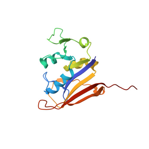
Crystal structure of Bacillus anthracis dihydrofolate reductase with the dihydrophthalazine-based trimethoprim derivative RAB1 provides a structural explanation of potency and selectivity.
Bourne, C.R., Bunce, R.A., Bourne, P.C., Berlin, K.D., Barrow, E.W., Barrow, W.W.(2009) Antimicrob Agents Chemother 53: 3065-3073
- PubMed: 19364848 Search on PubMedSearch on PubMed Central
- DOI: https://doi.org/10.1128/AAC.01666-08
- Primary Citation Related Structures:
3FL8, 3FL9 - PubMed Abstract:
Bacillus anthracis possesses an innate resistance to the antibiotic trimethoprim due to poor binding to dihydrofolate reductase (DHFR); currently, there are no commercial antibacterials that target this enzyme in B. anthracis. We have previously reported a series of dihydrophthalazine-based trimethoprim derivatives that are inhibitors for this target. In the present work, we have synthesized one compound (RAB1) displaying favorable 50% inhibitory concentration (54 nM) and MIC (< or =12.8 microg/ml) values. RAB1 was cocrystallized with the B. anthracis DHFR in the space group P2(1)2(1)2(1), and X-ray diffraction data were collected to a 2.3-A resolution. Binding of RAB1 causes a conformational change of the side chain of Arg58 and Met37 to accommodate the dihydrophthalazine moiety. Unlike the natural substrate or trimethoprim, the dihydrophthalazine group provides a large hydrophobic anchor that embeds within the DHFR active site and accounts for its selective inhibitory activity against B. anthracis.
- Department of Veterinary Pathobiology, Oklahoma State University, Stillwater, OK 74078, USA. christina.bourne@okstate.edu
Organizational Affiliation: