3FDL | pdb_00003fdl

Bim BH3 peptide in complex with Bcl-xL


Experimental Data Snapshot

  • Method: X-RAY DIFFRACTION
  • Resolution: 1.78 Å
  • R-Value Free: 
    0.208 (Depositor), 0.222 (DCC) 
  • R-Value Work: 
    0.160 (Depositor) 
  • R-Value Observed: 
    0.163 (Depositor) 

Starting Model: experimental
View more details

wwPDB Validation 3D Report Full Report

Validation slider image for 3FDL

This is version 1.4 of the entry. See complete history

Literature

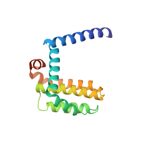
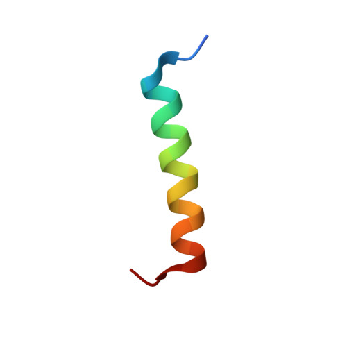
High-Resolution Structural Characterization of a Helical alpha/beta-Peptide Foldamer Bound to the Anti-Apoptotic Protein Bcl-x(L)

Lee, E.F.Sadowsky, J.D.Smith, B.J.Czabotar, P.E.Peterson-Kaufman, K.J.Colman, P.M.Gellman, S.H.Fairlie, W.D.

(2009) Angew Chem Int Ed Engl 48: 4318-4322

  • DOI: https://doi.org/10.1002/anie.200805761
  • Primary Citation Related Structures: 
    3FDL, 3FDM

  • PubMed Abstract: 

    Get into the groove: The first high-resolution structure of a foldamer bound to a protein target is described (see picture; foldamer in sticks). The foldamer consists of alpha- and beta-amino acid residues and is bound to the anti-apoptotic protein Bcl-x(L). The overall binding mode and key interactions observed in the foldamer/Bcl-x(L) complex mimic those seen in complexes of Bcl-x(L) with natural alpha-peptide ligands. Additional contacts in the foldamer/Bcl-x(L) complex involving beta-amino acid residues appear to contribute to binding affinity.


  • Organizational Affiliation
    • Structural Biology Division, The Walter and Eliza Hall Institute of Medical Research, 1G Royal Parade, Parkville, Victoria 3052, Australia.

Macromolecule Content 

  • Total Structure Weight: 21.19 kDa 
  • Atom Count: 1,594 
  • Modeled Residue Count: 168 
  • Deposited Residue Count: 184 
  • Unique protein chains: 2

Macromolecules

Find similar proteins by:|  3D Structure
Entity ID: 1
MoleculeChains  Sequence LengthOrganismDetailsImage
Apoptosis regulator Bcl-X158Homo sapiensMutation(s): 0 
Gene Names: Bcl-x
UniProt & NIH Common Fund Data Resources
Find proteins for Q07817 (Homo sapiens)
Explore Q07817 
Go to UniProtKB:  Q07817
PHAROS:  Q07817
GTEx:  ENSG00000171552 
Entity Groups
Sequence Clusters30% Identity50% Identity70% Identity90% Identity95% Identity100% Identity
UniProt GroupQ07817
Sequence Annotations
Expand
Reference Sequence
Find similar proteins by:|  3D Structure
Entity ID: 2
MoleculeChains  Sequence LengthOrganismDetailsImage
Bcl-2-like protein 1126synthetic constructMutation(s): 0 
UniProt & NIH Common Fund Data Resources
Find proteins for O43521 (Homo sapiens)
Explore O43521 
Go to UniProtKB:  O43521
PHAROS:  O43521
GTEx:  ENSG00000153094 
Entity Groups
Sequence Clusters30% Identity50% Identity70% Identity90% Identity95% Identity100% Identity
UniProt GroupO43521
Sequence Annotations
Expand
Reference Sequence

Experimental Data & Validation

Experimental Data

  • Method: X-RAY DIFFRACTION
  • Resolution: 1.78 Å
  • R-Value Free:  0.208 (Depositor), 0.222 (DCC) 
  • R-Value Work:  0.160 (Depositor) 
  • R-Value Observed: 0.163 (Depositor) 
Space Group: C 1 2 1
Unit Cell:
Length ( Å )Angle ( ˚ )
a = 74.111α = 90
b = 35.684β = 111.54
c = 62.76γ = 90
Software Package:
Software NamePurpose
CrystalCleardata collection
PHASERphasing
REFMACrefinement
HKL-2000data reduction
HKL-2000data scaling

Structure Validation

View Full Validation Report


Entry History 

Deposition Data

Revision History  (Full details and data files)

  • Version 1.0: 2009-03-10
    Type: Initial release
  • Version 1.1: 2011-07-13
    Changes: Version format compliance
  • Version 1.2: 2017-08-09
    Changes: Refinement description, Source and taxonomy
  • Version 1.3: 2017-08-16
    Changes: Source and taxonomy
  • Version 1.4: 2023-11-01
    Changes: Data collection, Database references, Refinement description