The MX2 macromolecular crystallography beamline: a wiggler X-ray source at the LNLS.
Guimaraes, B.G., Sanfelici, L., Neuenschwander, R.T., Rodrigues, F., Grizolli, W.C., Raulik, M.A., Piton, J.R., Meyer, B.C., Nascimento, A.S., Polikarpov, I.(2009) J Synchrotron Radiat 16: 69-75
- PubMed: 19096177
- DOI: https://doi.org/10.1107/S0909049508034870
- Primary Citation of Related Structures:
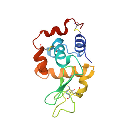
3EXD - PubMed Abstract:
The Brazilian Synchrotron Light Laboratory [Laboratório Nacional de Luz Síncrotron (LNLS), Campinas, SP, Brazil] is the first commissioned synchrotron light source in the southern hemisphere. The first wiggler macromolecular crystallography beamline (MX2) at the LNLS has been recently constructed and brought into operation. Here the technical design, experimental set-up, parameters of the beamline and the first experimental results obtained at MX2 are described. The beamline operates on a 2.0 T hybrid 30-pole wiggler, and its optical layout includes collimating mirror, Si(111) double-crystal monochromator and toroidal bendable mirror. The measured flux density at the sample position at 8.7 eV reaches 4.8 x 10(11) photons s(-1) mm(-2) (100 mA)(-1). The beamline is equipped with a MarResearch Desktop Beamline Goniostat (MarDTB) and 3 x 3 MarMosaic225 CCD detector, and is controlled by a customized version of the Blu-Ice software. A description of the first X-ray diffraction data sets collected at the MX2 LNLS beamline and used for macromolecular crystal structure solution is also provided.
- Laboratório Nacional de Luz Síncrotron (LNLS), Campinas, SP, Brazil. beatriz@lnls.br
Organizational Affiliation:


