3EU7

Crystal Structure of a PALB2 / BRCA2 complex


Experimental Data Snapshot

  • Method: X-RAY DIFFRACTION
  • Resolution: 2.20 Å
  • R-Value Free: 0.253 
  • R-Value Work: 0.197 
  • R-Value Observed: 0.200 

wwPDB Validation   3D Report Full Report


This is version 1.4 of the entry. See complete history


Literature

Structural basis for recruitment of BRCA2 by PALB2

Oliver, A.W.Swift, S.Lord, C.J.Ashworth, A.Pearl, L.H.

(2009) EMBO Rep 10: 990-996

  • DOI: https://doi.org/10.1038/embor.2009.126
  • Primary Citation of Related Structures:  
    2W18, 3EU7

  • PubMed Abstract: 

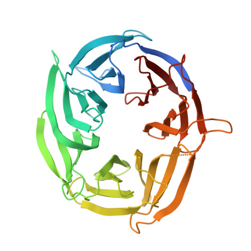
    The breast cancer 2, early onset protein (BRCA2) is central to the repair of DNA damage by homologous recombination. BRCA2 recruits the recombinase RAD51 to sites of damage, regulates its assembly into nucleoprotein filaments and thereby promotes homologous recombination. Localization of BRCA2 to nuclear foci requires its association with the partner and localizer of BRCA2 (PALB2), mutations in which are associated with cancer predisposition, as well as subtype N of Fanconi anaemia. We have determined the structure of the PALB2 carboxy-terminal beta-propeller domain in complex with a BRCA2 peptide. The structure shows the molecular determinants of this important protein-protein interaction and explains the effects of both cancer-associated truncating mutants in PALB2 and missense mutations in the amino-terminal region of BRCA2.


  • Organizational Affiliation

    Cancer Research UK DNA Repair Enzymes Group, Section of Structural Biology, 237 Fulham Road, London SW3 6JB, UK. antony.oliver@icr.ac.uk


Macromolecules
Find similar proteins by:  (by identity cutoff)  |  3D Structure
Entity ID: 1
MoleculeChains Sequence LengthOrganismDetailsImage
Partner and localizer of BRCA2356Homo sapiensMutation(s): 0 
Gene Names: PALB2
UniProt & NIH Common Fund Data Resources
Find proteins for Q86YC2 (Homo sapiens)
Explore Q86YC2 
Go to UniProtKB:  Q86YC2
PHAROS:  Q86YC2
GTEx:  ENSG00000083093 
Entity Groups  
Sequence Clusters30% Identity50% Identity70% Identity90% Identity95% Identity100% Identity
UniProt GroupQ86YC2
Sequence Annotations
Expand
  • Reference Sequence

Find similar proteins by:  Sequence   |   3D Structure  

Entity ID: 2
MoleculeChains Sequence LengthOrganismDetailsImage
19meric peptide from Breast cancer type 2 susceptibility proteinB [auth X]19N/AMutation(s): 0 
UniProt & NIH Common Fund Data Resources
Find proteins for P51587 (Homo sapiens)
Explore P51587 
Go to UniProtKB:  P51587
PHAROS:  P51587
GTEx:  ENSG00000139618 
Entity Groups  
Sequence Clusters30% Identity50% Identity70% Identity90% Identity95% Identity100% Identity
UniProt GroupP51587
Sequence Annotations
Expand
  • Reference Sequence
Small Molecules
Modified Residues  1 Unique
IDChains TypeFormula2D DiagramParent
CSD
Query on CSD
A
L-PEPTIDE LINKINGC3 H7 N O4 SCYS
Experimental Data & Validation

Experimental Data

  • Method: X-RAY DIFFRACTION
  • Resolution: 2.20 Å
  • R-Value Free: 0.253 
  • R-Value Work: 0.197 
  • R-Value Observed: 0.200 
  • Space Group: C 1 2 1
Unit Cell:
Length ( Å )Angle ( ˚ )
a = 82.815α = 90
b = 62.026β = 108.23
c = 77.975γ = 90
Software Package:
Software NamePurpose
PHENIXrefinement

Structure Validation

View Full Validation Report


Entry History 

Deposition Data

Revision History  (Full details and data files)

  • Version 1.0: 2009-07-28
    Type: Initial release
  • Version 1.1: 2011-07-13
    Changes: Non-polymer description, Version format compliance
  • Version 1.2: 2014-02-05
    Changes: Derived calculations
  • Version 1.3: 2014-11-12
    Changes: Other
  • Version 1.4: 2023-11-01
    Changes: Data collection, Database references, Derived calculations, Refinement description