Polysaccharide binding sites in hyaluronate lyase--crystal structures of native phage-encoded hyaluronate lyase and its complexes with ascorbic acid and lactose.
Mishra, P., Prem Kumar, R., Ethayathulla, A.S., Singh, N., Sharma, S., Perbandt, M., Betzel, C., Kaur, P., Srinivasan, A., Bhakuni, V., Singh, T.P.(2009) FEBS J 276: 3392-3402
- PubMed: 19438710 Search on PubMed
- DOI: https://doi.org/10.1111/j.1742-4658.2009.07065.x
- Primary Citation Related Structures:
2YW0, 3EKA - PubMed Abstract:
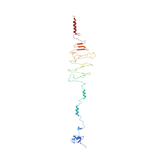
Hyaluronate lyases are a class of endoglycosaminidase enzymes with a high level of complexity and heterogeneity. The main function of the Streptococcus pyogenes bacteriophage protein hyaluronate lyase, HylP2, is to degrade hyaluronan into unsaturated disaccharide units. HylP2 was cloned, over-expressed and purified to homogeneity. The recombinant HylP2 exists as a homotrimer with a molecular mass of approximately 110 kDa under physiological conditions. The HylP2 was crystallized and the crystals were soaked in two separate reservoir solutions containing ascorbic acid and lactose, respectively. The crystal structures of native HylP2 and its two complexes with ascorbic acid and lactose have been determined. HylP2 folds into four distinct domains with a central core consisting of 16 antiparallel beta-strands forming an irregular triangular tube designated as triple-stranded beta-helix. The structures of complexes show that three molecules each of ascorbic acid and lactose bind to protein at the sugar binding groove in the triple-stranded beta-helix domain. Both ascorbic acid and lactose molecules occupy almost identical subsites in the long saccharide binding groove. Both ligands are involved in several hydrogen bonded interactions at each subsite. The binding characteristics and stereochemical properties indicate that Tyr264 may be involved in the catalytic activity of HylP2. The mutation of Tyr264 to Phe264 supports this observation.
- Department of Molecular and Structural Biology, Central Drug Research Institute, Lucknow, India.
Organizational Affiliation: