3EJH

Crystal Structure of the Fibronectin 8-9FnI Domain Pair in Complex with a Type-I Collagen Peptide


Experimental Data Snapshot

  • Method: X-RAY DIFFRACTION
  • Resolution: 2.10 Å
  • R-Value Free: 0.242 
  • R-Value Work: 0.209 
  • R-Value Observed: 0.211 

wwPDB Validation   3D Report Full Report


Ligand Structure Quality Assessment 


This is version 1.5 of the entry. See complete history


Literature

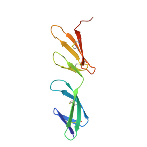
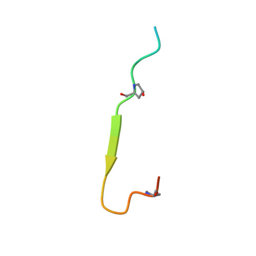
Identification and structural analysis of type I collagen sites in complex with fibronectin fragments.

Erat, M.C.Slatter, D.A.Lowe, E.D.Millard, C.J.Farndale, R.W.Campbell, I.D.Vakonakis, I.

(2009) Proc Natl Acad Sci U S A 106: 4195-4200

  • DOI: https://doi.org/10.1073/pnas.0812516106
  • Primary Citation of Related Structures:  
    3EJH

  • PubMed Abstract: 

    Collagen and fibronectin are major components of vertebrate extracellular matrices. Their association and distribution control the development and properties of diverse tissues, but thus far no structural information has been available for the complex formed. Here, we report binding of a peptide, derived from the alpha(1) chain of type I collagen, to the gelatin-binding domain of human fibronectin and present the crystal structure of this peptide in complex with the (8-9)FnI domain pair. Both gelatin-binding domain subfragments, (6)FnI(1-2)FnII(7)FnI and (8-9)FnI, bind the same specific sequence on D-period 4 of collagen I alpha(1), adjacent to the MMP-1 cleavage site. (8-9)FnI also binds the equivalent sequence of the alpha(2) chain. The collagen peptide adopts an antiparallel beta-strand conformation, similar to structures of proteins from pathogenic bacteria bound to FnI domains. Analysis of the type I collagen sequence suggests multiple putative fibronectin-binding sites compatible with our structural model. We demonstrate, by kinetic unfolding experiments, that the triple-helical collagen state is destabilized by (8-9)FnI. This finding suggests a role for fibronectin in collagen proteolysis and tissue remodeling.


  • Organizational Affiliation

    Department of Biochemistry, University of Oxford, Oxford OX1 3QU, United Kingdom.


Macromolecules
Find similar proteins by:  (by identity cutoff)  |  3D Structure
Entity ID: 1
MoleculeChains Sequence LengthOrganismDetailsImage
FibronectinA,
C [auth B]
93Homo sapiensMutation(s): 2 
Gene Names: FN1FN
UniProt & NIH Common Fund Data Resources
Find proteins for P02751 (Homo sapiens)
Explore P02751 
Go to UniProtKB:  P02751
PHAROS:  P02751
GTEx:  ENSG00000115414 
Entity Groups  
Sequence Clusters30% Identity50% Identity70% Identity90% Identity95% Identity100% Identity
UniProt GroupP02751
Sequence Annotations
Expand
  • Reference Sequence

Find similar proteins by:  Sequence   |   3D Structure  

Entity ID: 2
MoleculeChains Sequence LengthOrganismDetailsImage
Collagen type-I a1 chainB [auth E],
D [auth F]
23N/AMutation(s): 0 
UniProt & NIH Common Fund Data Resources
Find proteins for P02452 (Homo sapiens)
Explore P02452 
Go to UniProtKB:  P02452
PHAROS:  P02452
GTEx:  ENSG00000108821 
Entity Groups  
Sequence Clusters30% Identity50% Identity70% Identity90% Identity95% Identity100% Identity
UniProt GroupP02452
Sequence Annotations
Expand
  • Reference Sequence
Small Molecules
Modified Residues  1 Unique
IDChains TypeFormula2D DiagramParent
HYP
Query on HYP
B [auth E],
D [auth F]
L-PEPTIDE LINKINGC5 H9 N O3PRO
Experimental Data & Validation

Experimental Data

  • Method: X-RAY DIFFRACTION
  • Resolution: 2.10 Å
  • R-Value Free: 0.242 
  • R-Value Work: 0.209 
  • R-Value Observed: 0.211 
  • Space Group: P 31 2 1
Unit Cell:
Length ( Å )Angle ( ˚ )
a = 56.84α = 90
b = 56.84β = 90
c = 150.22γ = 120
Software Package:
Software NamePurpose
MOSFLMdata reduction
SCALAdata scaling
PHENIXrefinement
PDB_EXTRACTdata extraction
ADSCdata collection
PHASERphasing

Structure Validation

View Full Validation Report


Ligand Structure Quality Assessment 


Entry History 

Deposition Data

Revision History  (Full details and data files)

  • Version 1.0: 2009-02-03
    Type: Initial release
  • Version 1.1: 2011-07-13
    Changes: Non-polymer description, Version format compliance
  • Version 1.2: 2011-11-16
    Changes: Atomic model
  • Version 1.3: 2020-07-29
    Type: Remediation
    Reason: Carbohydrate remediation
    Changes: Advisory, Data collection, Derived calculations, Structure summary
  • Version 1.4: 2021-10-20
    Changes: Database references, Source and taxonomy, Structure summary
  • Version 1.5: 2023-08-30
    Changes: Data collection, Refinement description