3EIU

A second transient position of ATP on its trail to the nucleotide-binding site of subunit B of the motor protein A1Ao ATP synthase


Experimental Data Snapshot

  • Method: X-RAY DIFFRACTION
  • Resolution: 3.43 Å
  • R-Value Free: 0.315 
  • R-Value Work: 0.246 
  • R-Value Observed: 0.249 

wwPDB Validation   3D Report Full Report


Ligand Structure Quality Assessment 


This is version 1.4 of the entry. See complete history


Literature

A second transient position of ATP on its trail to the nucleotide-binding site of subunit B of the motor protein A(1)A(O) ATP synthase

Manimekalai, M.S.S.Kumar, A.Balakrishna, A.M.Gruber, G.

(2009) J Struct Biol 166: 38-45

  • DOI: https://doi.org/10.1016/j.jsb.2008.12.004

  • PubMed Abstract: 

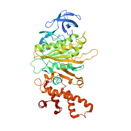
    The adenosine triphosphate (ATP) entrance into the nucleotide-binding subunits of ATP synthases is a puzzle. In the previously determined structure of subunit B mutant R416W of the Methanosarcina mazei Gö1 A-ATP synthase one ATP could be trapped at a transition position, close to the phosphate-binding loop. Using defined parameters for co-crystallization of an ATP-bound B-subunit, a unique transition position of ATP could be found in the crystallographic structure of this complex, solved at 3.4 A resolution. The nucleotide is found near the helix-turn-helix motif in the C-terminal domain of the protein; the location occupied by the gamma-subunit to interact with the empty beta-subunit in the thermoalkaliphilic Bacillus sp. TA2.A1 of the related F-ATP synthase. When compared with the determined structure of the ATP-transition position, close to the P-loop, and the nucleotide-free form of subunit B, the C-terminal domain of the B mutant is rotated by around 6 degrees, implicating an ATP moving pathway. We propose that, in the nucleotide empty state the central stalk subunit D is in close contact with subunit B and when the ATP molecule enters, D moves slightly, paving way for it to interact with the subunit B, which makes the C-terminal domain rotate by 6 degrees.


  • Organizational Affiliation

    Nanyang Technological University, Division of Structural & Computational Biology, School of Biological Sciences, 60 Nanyang Drive, Singapore 637551, Republic of Singapore.


Macromolecules
Find similar proteins by:  (by identity cutoff)  |  3D Structure
Entity ID: 1
MoleculeChains Sequence LengthOrganismDetailsImage
V-type ATP synthase beta chain
A, B
469Methanosarcina mazeiMutation(s): 1 
Gene Names: ATP Synthase
EC: 3.6.3.14
UniProt
Find proteins for Q60187 (Methanosarcina mazei (strain ATCC BAA-159 / DSM 3647 / Goe1 / Go1 / JCM 11833 / OCM 88))
Explore Q60187 
Go to UniProtKB:  Q60187
Entity Groups  
Sequence Clusters30% Identity50% Identity70% Identity90% Identity95% Identity100% Identity
UniProt GroupQ60187
Sequence Annotations
Expand
  • Reference Sequence
Small Molecules
Ligands 2 Unique
IDChains Name / Formula / InChI Key2D Diagram3D Interactions
ATP
Query on ATP

Download Ideal Coordinates CCD File 
C [auth A]ADENOSINE-5'-TRIPHOSPHATE
C10 H16 N5 O13 P3
ZKHQWZAMYRWXGA-KQYNXXCUSA-N
AES
Query on AES

Download Ideal Coordinates CCD File 
D [auth B]4-(2-AMINOETHYL)BENZENESULFONYL FLUORIDE
C8 H10 F N O2 S
MGSKVZWGBWPBTF-UHFFFAOYSA-N
Experimental Data & Validation

Experimental Data

  • Method: X-RAY DIFFRACTION
  • Resolution: 3.43 Å
  • R-Value Free: 0.315 
  • R-Value Work: 0.246 
  • R-Value Observed: 0.249 
  • Space Group: P 21 21 21
Unit Cell:
Length ( Å )Angle ( ˚ )
a = 75.693α = 90
b = 96.867β = 90
c = 129.631γ = 90
Software Package:
Software NamePurpose
DENZOdata reduction
SCALEPACKdata scaling
MOLREPphasing
REFMACrefinement
PDB_EXTRACTdata extraction
ADSCdata collection
HKL-2000data reduction
HKL-2000data scaling

Structure Validation

View Full Validation Report


Ligand Structure Quality Assessment 


Entry History 

Deposition Data

Revision History  (Full details and data files)

  • Version 1.0: 2009-02-10
    Type: Initial release
  • Version 1.1: 2011-07-13
    Changes: Advisory, Version format compliance
  • Version 1.2: 2017-10-25
    Changes: Refinement description
  • Version 1.3: 2021-11-10
    Changes: Database references, Derived calculations
  • Version 1.4: 2023-11-01
    Changes: Data collection, Refinement description