3EG5 | pdb_00003eg5

Crystal structure of MDIA1-TSH GBD-FH3 in complex with CDC42-GMPPNP


Experimental Data Snapshot

  • Method: X-RAY DIFFRACTION
  • Resolution: 2.70 Å
  • R-Value Free: 
    0.243 (Depositor), 0.231 (DCC) 
  • R-Value Work: 
    0.206 (Depositor), 0.215 (DCC) 
  • R-Value Observed: 
    0.208 (Depositor) 

Starting Model: experimental
View more details

wwPDB Validation 3D Report Full Report

Validation slider image for 3EG5

Ligand Structure Quality Assessment 


This is version 1.3 of the entry. See complete history

Literature

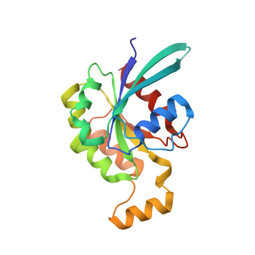
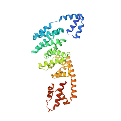
Specificity of Interactions between mDia Isoforms and Rho Proteins

Lammers, M.Meyer, S.Kuhlmann, D.Wittinghofer, A.

(2008) J Biological Chem 283: 35236-35246

  • DOI: https://doi.org/10.1074/jbc.M805634200
  • Primary Citation Related Structures: 
    3EG5

  • PubMed Abstract: 

    Formins are key regulators of actin nucleation and polymerization. They contain formin homology 1 (FH1) and 2 (FH2) domains as the catalytic machinery for the formation of linear actin cables. A subclass of formins constitutes the Diaphanous-related formins, members of which are regulated by the binding of a small GTP-binding protein of the Rho subfamily. Binding of these molecular switch proteins to the regulatory N-terminal mDia(N), including the GTPase-binding domain, leads to the release of auto-inhibition. From the three mDia isoforms, mDia1 is activated only by Rho (RhoA, -B, and -C), in contrast to mDia2 and -3, which is also activated by Rac and Cdc42. Little is known about the determinants of specificity. Here we report on the interactions of RhoA, Rac1, and Cdc42 with mDia1 and an mDia1 mutant (mDia(N)-Thr-Ser-His (TSH)), which based on structural information should mimic mDia2 and -3. Specificity is analyzed by biochemical studies and a structural analysis of a complex between Cdc42.Gpp(NH)p and mDia(N)-TSH. A triple NNN motif in mDia1 (amino acids 164-166), corresponding to the TSH motif in mDia2/3 (amino acids 183-185 and 190-192), and the epitope interacting with the Rho insert helix are essential for high affinity binding. The triple N motif of mDia1 allows tight interaction with Rho because of the presence of Phe-106, whereas the corresponding His-104 in Rac and Cdc42 forms a complementary interface with the TSH motif in mDia2/3. We also show that the F106H and H104F mutations drastically alter the affinities and thermodynamics of mDia interactions.


  • Organizational Affiliation
    • Max-Planck-Institute for Molecular Physiology, Department of Structural Biology, Otto-Hahn-Strasse 11, 44227 Dortmund, Germany.

Macromolecule Content 

  • Total Structure Weight: 129.1 kDa 
  • Atom Count: 8,341 
  • Modeled Residue Count: 1,030 
  • Deposited Residue Count: 1,122 
  • Unique protein chains: 2

Macromolecules

Find similar proteins by:|  3D Structure
Entity ID: 1
MoleculeChains  Sequence LengthOrganismDetailsImage
Cell division control protein 42 homolog
A, C
178Mus musculusMutation(s): 0 
Gene Names: Cdc42
EC: 3.6.5.2
UniProt & NIH Common Fund Data Resources
Find proteins for P60766 (Mus musculus)
Explore P60766 
Go to UniProtKB:  P60766
IMPC:  MGI:106211
Entity Groups
Sequence Clusters30% Identity50% Identity70% Identity90% Identity95% Identity100% Identity
UniProt GroupP60766
Sequence Annotations
Expand
Reference Sequence
Find similar proteins by:|  3D Structure
Entity ID: 2
MoleculeChains  Sequence LengthOrganismDetailsImage
Protein diaphanous homolog 1
B, D
383Mus musculusMutation(s): 3 
Gene Names: Diaph1
UniProt & NIH Common Fund Data Resources
Find proteins for O08808 (Mus musculus)
Explore O08808 
Go to UniProtKB:  O08808
IMPC:  MGI:1194490
Entity Groups
Sequence Clusters30% Identity50% Identity70% Identity90% Identity95% Identity100% Identity
UniProt GroupO08808
Sequence Annotations
Expand
Reference Sequence

Experimental Data & Validation

Experimental Data

  • Method: X-RAY DIFFRACTION
  • Resolution: 2.70 Å
  • R-Value Free:  0.243 (Depositor), 0.231 (DCC) 
  • R-Value Work:  0.206 (Depositor), 0.215 (DCC) 
  • R-Value Observed: 0.208 (Depositor) 
Space Group: P 32
Unit Cell:
Length ( Å )Angle ( ˚ )
a = 71.269α = 90
b = 71.269β = 90
c = 244.964γ = 120
Software Package:
Software NamePurpose
MOLREPphasing
REFMACrefinement
XDSdata reduction
XDSdata scaling

Structure Validation

View Full Validation Report


Ligand Structure Quality Assessment 


Entry History 

Deposition Data

Revision History  (Full details and data files)

  • Version 1.0: 2008-10-14
    Type: Initial release
  • Version 1.1: 2011-07-13
    Changes: Version format compliance
  • Version 1.2: 2021-11-10
    Changes: Database references, Derived calculations
  • Version 1.3: 2023-11-01
    Changes: Data collection, Refinement description