3EFU | pdb_00003efu

X-ray structure of human ubiquitin-Hg(II) adduct


Experimental Data Snapshot

  • Method: X-RAY DIFFRACTION
  • Resolution: 1.84 Å
  • R-Value Free: 
    0.293 (Depositor), 0.303 (DCC) 
  • R-Value Work: 
    0.235 (Depositor), 0.240 (DCC) 
  • R-Value Observed: 
    0.235 (Depositor) 

Starting Model: experimental
View more details

wwPDB Validation 3D Report Full Report

Validation slider image for 3EFU

This is version 1.2 of the entry. See complete history

Literature

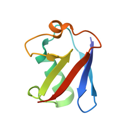
Structural probing of Zn(ii), Cd(ii) and Hg(ii) binding to human ubiquitin.

Falini, G.Fermani, S.Tosi, G.Arnesano, F.Natile, G.

(2008) Chem Commun (Camb) 45: 5960-5962

  • DOI: https://doi.org/10.1039/b813463d
  • Primary Citation Related Structures: 
    3EEC, 3EFU, 3EHV

  • PubMed Abstract: 

    A structural investigation performed on adducts of human ubiquitin with group-12 metal ions reveals common preferential anchoring sites, the most populated one being His68; at higher metal ion concentration a second and a third site, close to the N-terminus of the protein, become populated and promote a polymorphic transition from orthorhombic to cubic form; Glu16 and Glu18, involved in the latter metal binding, undergo a remarkable displacement from their position in native ubiquitin; the aggregate stereochemistry appears to be driven by the clustering of deshielded backbone hydrogen-bond patches, and metal ions foster this process.


  • Organizational Affiliation
    • Dipartimento di Chimica, G. Ciamician, Università di Bologna, via Selmi 2, 40126, Bologna, Italy. giuseppe.falini@unibo.it

Macromolecule Content 

  • Total Structure Weight: 8.78 kDa 
  • Atom Count: 693 
  • Modeled Residue Count: 72 
  • Deposited Residue Count: 76 
  • Unique protein chains: 1

Macromolecules

Find similar proteins by:|  3D Structure
Entity ID: 1
MoleculeChains  Sequence LengthOrganismDetailsImage
Ubiquitin76Homo sapiensMutation(s): 0 
Gene Names: RPS27AUBA80UBCEP1UBA52UBCEP2UBBUBC
UniProt & NIH Common Fund Data Resources
Find proteins for P0CG48 (Homo sapiens)
Explore P0CG48 
Go to UniProtKB:  P0CG48
PHAROS:  P0CG48
GTEx:  ENSG00000150991 
Entity Groups
Sequence Clusters30% Identity50% Identity70% Identity90% Identity95% Identity100% Identity
UniProt GroupP0CG48
Sequence Annotations
Expand
Reference Sequence

Small Molecules

Ligands 1 Unique
IDChains Name / Formula / InChI Key2D Diagram3D Interactions
HG

Query on HG


Download:Ideal Coordinates CCD File
B [auth A]MERCURY (II) ION
Hg
BQPIGGFYSBELGY-UHFFFAOYSA-N

Experimental Data & Validation

Experimental Data

  • Method: X-RAY DIFFRACTION
  • Resolution: 1.84 Å
  • R-Value Free:  0.293 (Depositor), 0.303 (DCC) 
  • R-Value Work:  0.235 (Depositor), 0.240 (DCC) 
  • R-Value Observed: 0.235 (Depositor) 
Space Group: P 21 21 21
Unit Cell:
Length ( Å )Angle ( ˚ )
a = 28.024α = 90
b = 42.828β = 90
c = 50.374γ = 90
Software Package:
Software NamePurpose
AMoREphasing
CNSrefinement
DENZOdata reduction
SCALEPACKdata scaling

Structure Validation

View Full Validation Report


Entry History 

Deposition Data

Revision History  (Full details and data files)

  • Version 1.0: 2008-12-09
    Type: Initial release
  • Version 1.1: 2011-07-13
    Changes: Version format compliance
  • Version 1.2: 2023-11-01
    Changes: Data collection, Database references, Derived calculations, Refinement description