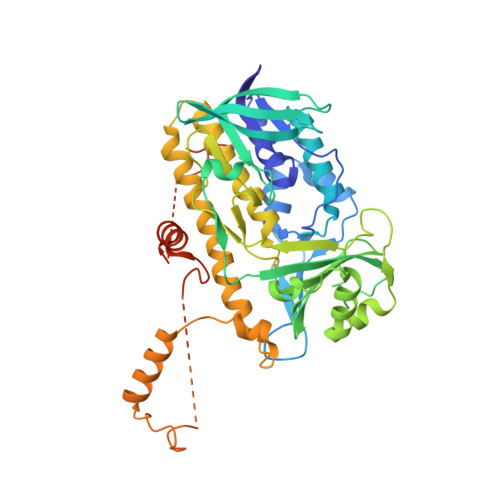
Structure and action of the myxobacterial chondrochloren halogenase CndH: a new variant of FAD-dependent halogenases.
Buedenbender, S., Rachid, S., Muller, R., Schulz, G.E.(2009) J Mol Biology 385: 520-530
- PubMed: 19000696
- DOI: https://doi.org/10.1016/j.jmb.2008.10.057
- Primary Citation of Related Structures:
3E1T - PubMed Abstract:
The crystal structure of the FAD-dependent chondrochloren halogenase CndH has been established at 2.1 A resolution. The enzyme contains the characteristic FAD-binding scaffold of the glutathione reductase superfamily. Except for its C-terminal domain, the chainfold of CndH is virtually identical with those of FAD-dependent aromatic hydroxylases. When compared to the structurally known FAD-dependent halogenases PrnA and RebH, CndH lacks a 45 residue segment near position 100 and deviates in the C-terminal domain. Both variations are near the active center and appear to reflect substrate differences. Whereas PrnA and RebH modify free tryptophan, CndH halogenates the tyrosyl group of a chondrochloren precursor that is most likely bound to a carrier protein. In contrast to PrnA and RebH, which enclose their small substrate completely, CndH has a large non-polar surface patch that may accommodate the putative carrier. Apart from the substrate binding site, the active center of CndH corresponds to those of PrnA and RebH. At the halogenation site, CndH has the characteristic lysine (Lys76) but lacks the required base Glu346 (PrnA). This base may be supplied by a residue of its C-terminal domain or by the carrier. These differences were corroborated by an overall sequence comparison between the known FAD-dependent halogenases, which revealed a split into a PrnA-RebH group and a CndH group. The two functionally established members of the CndH group use carrier-bound substrates, whereas three members of PrnA-RebH group are known to accept a free amino acid. Given the structural and functional distinction, we classify CndH as a new variant B of the FAD-dependent halogenases, adding a new feature to the structurally established variant A enzymes PrnA and RebH.
- Institut für Organische Chemie und Biochemie, Albert-Ludwigs-Universität, Albertstr. 21, D-79104 Freiburg im Breisgau, Germany.
Organizational Affiliation:


