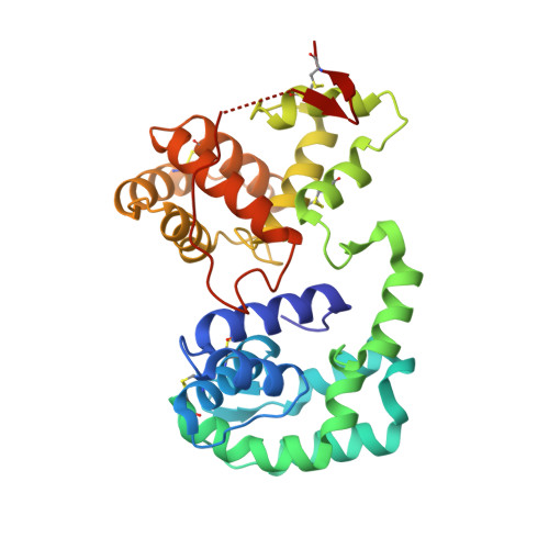
3DZT

AeD7-leukotriene E4 complex


Experimental Data Snapshot

  • Method: X-RAY DIFFRACTION
  • Resolution: 1.80 Å
  • R-Value Free: 0.201 
  • R-Value Work: 0.160 
  • R-Value Observed: 0.162 

wwPDB Validation   3D Report Full Report


Ligand Structure Quality Assessment 


This is version 1.3 of the entry. See complete history


Literature

Multifunctionality and mechanism of ligand binding in a mosquito antiinflammatory protein

Calvo, E.Mans, B.J.Ribeiro, J.M.Andersen, J.F.

(2009) Proc Natl Acad Sci U S A 106: 3728-3733

  • DOI: 10.1073/pnas.0813190106
  • Primary Citation of Related Structures:  
    3DXL, 3DY9, 3DYE, 3DZT

  • PubMed Abstract: 
  • The mosquito D7 salivary proteins are encoded by a multigene family related to the arthropod odorant-binding protein (OBP) superfamily. Forms having either one or two OBP domains are found in mosquito saliva. Four single-domain and one two-domain D7 proteins from Anopheles gambiae and Aedes aegypti (AeD7), respectively, were shown to bind biogenic amines with high affinity and with a stoichiometry of one ligand per protein molecule ...

    The mosquito D7 salivary proteins are encoded by a multigene family related to the arthropod odorant-binding protein (OBP) superfamily. Forms having either one or two OBP domains are found in mosquito saliva. Four single-domain and one two-domain D7 proteins from Anopheles gambiae and Aedes aegypti (AeD7), respectively, were shown to bind biogenic amines with high affinity and with a stoichiometry of one ligand per protein molecule. Sequence comparisons indicated that only the C-terminal domain of AeD7 is homologous to the single-domain proteins from A. gambiae, suggesting that the N-terminal domain may bind a different class of ligands. Here, we describe the 3D structure of AeD7 and examine the ligand-binding characteristics of the N- and C-terminal domains. Isothermal titration calorimetry and ligand complex crystal structures show that the N-terminal domain binds cysteinyl leukotrienes (cysLTs) with high affinities (50-60 nM) whereas the C-terminal domain binds biogenic amines. The lipid chain of the cysLT binds in a hydrophobic pocket of the N-terminal domain, whereas binding of norepinephrine leads to an ordering of the C-terminal portion of the C-terminal domain into an alpha-helix that, along with rotations of Arg-176 and Glu-268 side chains, acts to bury the bound ligand.


    Organizational Affiliation

    Laboratory of Malaria and Vector Research, National Institute of Allergy and Infectious Diseases, National Institutes of Health, Rockville, MD 20852, USA.


Macromolecules
Find similar proteins by:  (by identity cutoff)  |  3D Structure
Entity ID: 1
MoleculeChainsSequence LengthOrganismDetailsImage
D7 proteinA303Aedes aegyptiMutation(s): 2 
Gene Names: D7AAEL006424
UniProt
Find proteins for P18153 (Aedes aegypti)
Explore P18153 
Go to UniProtKB:  P18153
Entity Groups  
Sequence Clusters30% Identity50% Identity70% Identity90% Identity95% Identity100% Identity
UniProt GroupP18153
Protein Feature View
Expand
  • Reference Sequence
Small Molecules
Ligands 4 Unique
IDChainsName / Formula / InChI Key2D Diagram3D Interactions
EAH
Query on EAH

Download Ideal Coordinates CCD File 
C [auth A](5S,7E,9E,11Z,14Z)-5-hydroxyicosa-7,9,11,14-tetraenoic acid
C20 H32 O3
DVGDBDLZYCUGNU-LBIUVHPXSA-N
 Ligand Interaction
TRS
Query on TRS

Download Ideal Coordinates CCD File 
D [auth A]2-AMINO-2-HYDROXYMETHYL-PROPANE-1,3-DIOL
C4 H12 N O3
LENZDBCJOHFCAS-UHFFFAOYSA-O
 Ligand Interaction
GOL
Query on GOL

Download Ideal Coordinates CCD File 
E [auth A],
F [auth A],
G [auth A]
GLYCEROL
C3 H8 O3
PEDCQBHIVMGVHV-UHFFFAOYSA-N
 Ligand Interaction
CL
Query on CL

Download Ideal Coordinates CCD File 
B [auth A]CHLORIDE ION
Cl
VEXZGXHMUGYJMC-UHFFFAOYSA-M
 Ligand Interaction
Binding Affinity Annotations 
IDSourceBinding Affinity
EAH PDBBind:  3DZT Kd: 60.2 (nM) from 1 assay(s)
Binding MOAD:  3DZT Kd: 60.2 (nM) from 1 assay(s)
Experimental Data & Validation

Experimental Data

  • Method: X-RAY DIFFRACTION
  • Resolution: 1.80 Å
  • R-Value Free: 0.201 
  • R-Value Work: 0.160 
  • R-Value Observed: 0.162 
  • Space Group: P 1 21 1
Unit Cell:
Length ( Å )Angle ( ˚ )
a = 49.567α = 90
b = 65.307β = 112.34
c = 52.195γ = 90
Software Package:
Software NamePurpose
DENZOdata reduction
SCALEPACKdata scaling
REFMACrefinement
PDB_EXTRACTdata extraction
MAR345dtbdata collection
FFTphasing

Structure Validation

View Full Validation Report


Ligand Structure Quality Assessment 


Entry History 

Deposition Data

Revision History  (Full details and data files)

  • Version 1.0: 2009-02-03
    Type: Initial release
  • Version 1.1: 2011-07-13
    Changes: Advisory, Version format compliance
  • Version 1.2: 2017-10-25
    Changes: Refinement description
  • Version 1.3: 2021-03-31
    Changes: Derived calculations, Source and taxonomy