3DSR

ADP in transition binding site in the subunit B of the energy converter A1Ao ATP synthase


Experimental Data Snapshot

  • Method: X-RAY DIFFRACTION
  • Resolution: 2.70 Å
  • R-Value Free: 0.267 
  • R-Value Work: 0.198 
  • R-Value Observed: 0.201 

wwPDB Validation   3D Report Full Report


Ligand Structure Quality Assessment 


This is version 1.2 of the entry. See complete history


Literature

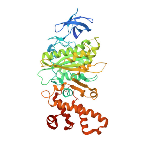
Structure of the nucleotide-binding subunit B of the energy producer A1A0 ATP synthase in complex with adenosine diphosphate

Kumar, A.Manimekalai, M.S.Gruber, G.

(2008) Acta Crystallogr D Biol Crystallogr 64: 1110-1115

  • DOI: https://doi.org/10.1107/S090744490802790X
  • Primary Citation of Related Structures:  
    3DSR

  • PubMed Abstract: 

    A1A0 ATP synthases are the major energy producers in archaea. Like the related prokaryotic and eukaryotic F1F0 ATP synthases, they are responsible for most of the synthesis of adenosine triphosphate. The catalytic events of A1A0 ATP synthases take place inside the A3B3 hexamer of the A1 domain. Recently, the crystallographic structure of the nucleotide-free subunit B of Methanosarcina mazei Gö1 A1A0 ATP synthase has been determined at 1.5 A resolution. To understand more about the nucleotide-binding mechanism, a protocol has been developed to crystallize the subunit B-ADP complex. The crystallographic structure of this complex has been solved at 2.7 A resolution. The ADP occupies a position between the essential phosphate-binding loop and amino-acid residue Phe149, which are involved in the binding of the antibiotic efrapeptin in the related F1F0 ATP synthases. This trapped ADP location is about 13 A distant from its final binding site and is therefore called the transition ADP-binding position. In the trapped ADP position the structure of subunit B adopts a different conformation, mainly in its C-terminal domain and also in the final nucleotide-binding site of the central alphabeta-domain. This atomic model provides insight into how the substrate enters into the nucleotide-binding protein and thereby into the catalytic A3B3 domain.


  • Organizational Affiliation

    Nanyang Technological University, School of Biological Sciences, 60 Nanyang Drive, Singapore 637551, Singapore.


Macromolecules
Find similar proteins by:  (by identity cutoff)  |  3D Structure
Entity ID: 1
MoleculeChains Sequence LengthOrganismDetailsImage
V-type ATP synthase beta chain
A, B
469Methanosarcina mazeiMutation(s): 0 
Gene Names: ATP Synthase
EC: 3.6.3.14
UniProt
Find proteins for Q60187 (Methanosarcina mazei (strain ATCC BAA-159 / DSM 3647 / Goe1 / Go1 / JCM 11833 / OCM 88))
Explore Q60187 
Go to UniProtKB:  Q60187
Entity Groups  
Sequence Clusters30% Identity50% Identity70% Identity90% Identity95% Identity100% Identity
UniProt GroupQ60187
Sequence Annotations
Expand
  • Reference Sequence
Small Molecules
Ligands 1 Unique
IDChains Name / Formula / InChI Key2D Diagram3D Interactions
ADP
Query on ADP

Download Ideal Coordinates CCD File 
C [auth A]ADENOSINE-5'-DIPHOSPHATE
C10 H15 N5 O10 P2
XTWYTFMLZFPYCI-KQYNXXCUSA-N
Experimental Data & Validation

Experimental Data

  • Method: X-RAY DIFFRACTION
  • Resolution: 2.70 Å
  • R-Value Free: 0.267 
  • R-Value Work: 0.198 
  • R-Value Observed: 0.201 
  • Space Group: P 21 21 21
Unit Cell:
Length ( Å )Angle ( ˚ )
a = 74.209α = 90
b = 95.944β = 90
c = 130.38γ = 90
Software Package:
Software NamePurpose
MOLREPphasing
REFMACrefinement
PDB_EXTRACTdata extraction
ADSCdata collection
HKL-2000data reduction
HKL-2000data scaling

Structure Validation

View Full Validation Report


Ligand Structure Quality Assessment 


Entry History 

Deposition Data

Revision History  (Full details and data files)

  • Version 1.0: 2009-06-16
    Type: Initial release
  • Version 1.1: 2011-07-13
    Changes: Advisory, Version format compliance
  • Version 1.2: 2023-11-01
    Changes: Data collection, Database references, Derived calculations, Refinement description