3DGT

The 1.5 A crystal structure of endo-1,3-beta-glucanase from Streptomyces sioyaensis


Experimental Data Snapshot

  • Method: X-RAY DIFFRACTION
  • Resolution: 1.50 Å
  • R-Value Free: 0.198 
  • R-Value Work: 0.183 
  • R-Value Observed: 0.184 

wwPDB Validation   3D Report Full Report


This is version 1.2 of the entry. See complete history


Literature

The 1.5 A structure of endo-1,3-beta-glucanase from Streptomyces sioyaensis: evolution of the active-site structure for 1,3-beta-glucan-binding specificity and hydrolysis

Hong, T.-Y.Hsiao, Y.-Y.Meng, M.Li, T.T.

(2008) Acta Crystallogr D Biol Crystallogr 64: 964-970

  • DOI: https://doi.org/10.1107/S0907444908021550
  • Primary Citation of Related Structures:  
    3DGT

  • PubMed Abstract: 

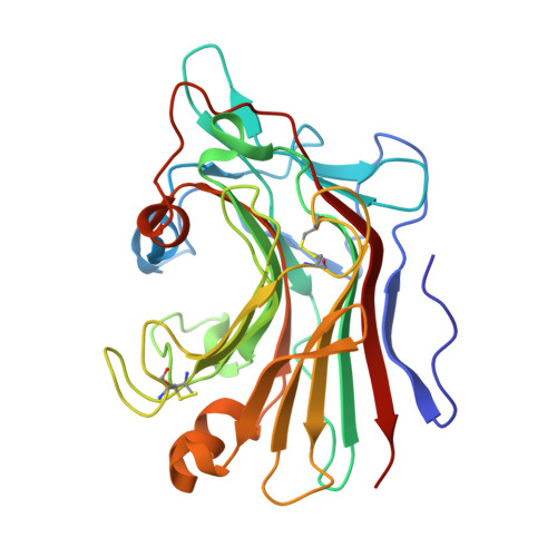
    The catalytic domain structure of Streptomyces sioyaensis 1,3-beta-glucanase (278 amino acids), a member of glycosyl hydrolase family 16 (GHF16), was determined to 1.5 A resolution in space group P2(1)2(1)2(1). The enzyme specifically hydrolyzes the glycosidic bond of the 1,3-beta-linked glucan substrate. The overall structure contains two antiparallel six-and seven-stranded beta-sheets stacked in a beta-sandwich jelly-roll motif similar to the fold of GHF16 1,3-1,4-beta-glucanases. The active-site cleft of the enzyme is distinct, with the closure of one end primarily caused by two protruding loop insertions and two key residues, Tyr38 and Tyr134. The current known structures of 1,3-1,4-beta-glucanases and 1,3-beta-glucanase from Nocardiopsis sp., on the other hand, have open-channel active-site clefts that can accommodate six beta-D-glucopyranosyl units. The active-site structure of 1,3-beta-glucanase was compared with those of other homologous structures in order to address the binding and enzymatic specificity for 1,3-beta-linked glucans in Streptomyces. This information could be helpful in the development of specific antifungal agents.


  • Organizational Affiliation

    Department of Biotechnology, Tajen University, Pingtung 907, Taiwan.


Macromolecules
Find similar proteins by:  (by identity cutoff)  |  3D Structure
Entity ID: 1
MoleculeChains Sequence LengthOrganismDetailsImage
Endo-1,3-beta-glucanase280Streptomyces sioyaensisMutation(s): 0 
EC: 3.2.1.39
UniProt
Find proteins for Q9L816 (Streptomyces sioyaensis)
Explore Q9L816 
Go to UniProtKB:  Q9L816
Entity Groups  
Sequence Clusters30% Identity50% Identity70% Identity90% Identity95% Identity100% Identity
UniProt GroupQ9L816
Sequence Annotations
Expand
  • Reference Sequence
Small Molecules
Ligands 1 Unique
IDChains Name / Formula / InChI Key2D Diagram3D Interactions
MG
Query on MG

Download Ideal Coordinates CCD File 
B [auth A]MAGNESIUM ION
Mg
JLVVSXFLKOJNIY-UHFFFAOYSA-N
Experimental Data & Validation

Experimental Data

  • Method: X-RAY DIFFRACTION
  • Resolution: 1.50 Å
  • R-Value Free: 0.198 
  • R-Value Work: 0.183 
  • R-Value Observed: 0.184 
  • Space Group: P 21 21 21
Unit Cell:
Length ( Å )Angle ( ˚ )
a = 39.509α = 90
b = 75.967β = 90
c = 79.663γ = 90
Software Package:
Software NamePurpose
REFMACrefinement
CrystalCleardata collection
DENZOdata reduction
SCALEPACKdata scaling
SHELXCDphasing
SHELXEmodel building

Structure Validation

View Full Validation Report


Entry History 

Deposition Data

  • Released Date: 2008-09-02 
  • Deposition Author(s): Li, T.H.

Revision History  (Full details and data files)

  • Version 1.0: 2008-09-02
    Type: Initial release
  • Version 1.1: 2011-07-13
    Changes: Version format compliance
  • Version 1.2: 2017-10-25
    Changes: Refinement description