3DEQ

Crystal structure of dipeptide epimerase from Thermotoga maritima complexed with L-Ala-L-Leu dipeptide


Experimental Data Snapshot

  • Method: X-RAY DIFFRACTION
  • Resolution: 2.10 Å
  • R-Value Free: 0.259 
  • R-Value Work: 0.246 
  • R-Value Observed: 0.246 

wwPDB Validation   3D Report Full Report


This is version 1.3 of the entry. See complete history


Literature

Discovery of a dipeptide epimerase enzymatic function guided by homology modeling and virtual screening.

Kalyanaraman, C.Imker, H.J.Fedorov, A.A.Fedorov, E.V.Glasner, M.E.Babbitt, P.C.Almo, S.C.Gerlt, J.A.Jacobson, M.P.

(2008) Structure 16: 1668-1677

  • DOI: https://doi.org/10.1016/j.str.2008.08.015
  • Primary Citation of Related Structures:  
    3DEQ, 3DER, 3DES, 3DFY

  • PubMed Abstract: 

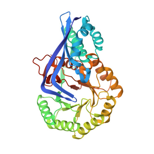
    We have developed a computational approach to aid the assignment of enzymatic function for uncharacterized proteins that uses homology modeling to predict the structure of the binding site and in silico docking to identify potential substrates. We apply this method to proteins in the functionally diverse enolase superfamily that are homologous to the characterized L-Ala-D/L-Glu epimerase from Bacillus subtilis. In particular, a protein from Thermotoga martima was predicted to have different substrate specificity, which suggests that it has a different, but as yet unknown, biological function. This prediction was experimentally confirmed, resulting in the assignment of epimerase activity for L-Ala-D/L-Phe, L-Ala-D/L-Tyr, and L-Ala-D/L-His, whereas the enzyme is annotated incorrectly in GenBank as muconate cycloisomerase. Subsequently, crystal structures of the enzyme were determined in complex with three substrates, showing close agreement with the computational models and revealing the structural basis for the observed substrate selectivity.


  • Organizational Affiliation

    Department of Pharmaceutical Chemistry, School of Pharmacy, University of California, 600 16(th) Street, San Francisco, CA 94158, USA.


Macromolecules
Find similar proteins by:  (by identity cutoff)  |  3D Structure
Entity ID: 1
MoleculeChains Sequence LengthOrganismDetailsImage
Muconate cycloisomerase
A, B, C, D
345Thermotoga maritima MSB8Mutation(s): 0 
Gene Names: TM_0006
UniProt
Find proteins for Q9WXM1 (Thermotoga maritima (strain ATCC 43589 / DSM 3109 / JCM 10099 / NBRC 100826 / MSB8))
Explore Q9WXM1 
Go to UniProtKB:  Q9WXM1
Entity Groups  
Sequence Clusters30% Identity50% Identity70% Identity90% Identity95% Identity100% Identity
UniProt GroupQ9WXM1
Sequence Annotations
Expand
  • Reference Sequence
Small Molecules
Ligands 3 Unique
IDChains Name / Formula / InChI Key2D Diagram3D Interactions
LEU
Query on LEU

Download Ideal Coordinates CCD File 
G [auth A],
J [auth B],
M [auth C],
P [auth D]
LEUCINE
C6 H13 N O2
ROHFNLRQFUQHCH-YFKPBYRVSA-N
ALA
Query on ALA

Download Ideal Coordinates CCD File 
F [auth A],
I [auth B],
L [auth C],
O [auth D]
ALANINE
C3 H7 N O2
QNAYBMKLOCPYGJ-REOHCLBHSA-N
MG
Query on MG

Download Ideal Coordinates CCD File 
E [auth A],
H [auth B],
K [auth C],
N [auth D]
MAGNESIUM ION
Mg
JLVVSXFLKOJNIY-UHFFFAOYSA-N
Experimental Data & Validation

Experimental Data

  • Method: X-RAY DIFFRACTION
  • Resolution: 2.10 Å
  • R-Value Free: 0.259 
  • R-Value Work: 0.246 
  • R-Value Observed: 0.246 
  • Space Group: P 61 2 2
Unit Cell:
Length ( Å )Angle ( ˚ )
a = 191.581α = 90
b = 191.581β = 90
c = 283.234γ = 120
Software Package:
Software NamePurpose
CNSrefinement
ADSCdata collection
DENZOdata reduction
SCALEPACKdata scaling
BALBESphasing

Structure Validation

View Full Validation Report


Entry History 

Deposition Data

Revision History  (Full details and data files)

  • Version 1.0: 2008-11-25
    Type: Initial release
  • Version 1.1: 2011-07-13
    Changes: Version format compliance
  • Version 1.2: 2023-08-30
    Changes: Data collection, Database references, Derived calculations, Refinement description
  • Version 1.3: 2024-03-13
    Changes: Source and taxonomy, Structure summary