Re-engineering specificity in 1,3-1,4-beta-glucanase to accept branched xyloglucan substrates
Addington, T., Calisto, B., Alfonso-Prieto, M., Rovira, C., Fita, I., Planas, A.(2011) Proteins 79: 365-375
- PubMed: 21069723 Search on PubMed
- DOI: https://doi.org/10.1002/prot.22884
- Primary Citation Related Structures:
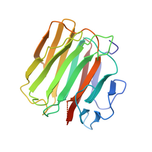
3D6E - PubMed Abstract:
Family 16 carbohydrate active enzyme members Bacillus licheniformis 1,3-1,4-β-glucanase and Populus tremula x tremuloides xyloglucan endotransglycosylase (XET16-34) are highly structurally related but display different substrate specificities. Although the first binds linear gluco-oligosaccharides, the second binds branched xylogluco-oligosaccharides. Prior engineered nucleophile mutants of both enzymes are glycosynthases that catalyze the condensation between a glycosyl fluoride donor and a glycoside acceptor. With the aim of expanding the glycosynthase technology to produce designer oligosaccharides consisting of hybrids between branched xylogluco- and linear gluco-oligosaccharides, enzyme engineering on the negative subsites of 1,3-1,4-β-glucanase to accept branched substrates has been undertaken. Removal of the 1,3-1,4-β-glucanase major loop and replacement with that of XET16-34 to open the binding cleft resulted in a folded protein, which still maintained some β-glucan hydrolase activity, but the corresponding nucleophile mutant did not display glycosynthase activity with either linear or branched glycosyl donors. Next, point mutations of the 1,3-1,4-β-glucanase β-sheets forming the binding site cleft were mutated to resemble XET16-34 residues. The final chimeric protein acquired binding affinity for xyloglucan and did not bind β-glucan. Therefore, binding specificity has been re-engineered, but affinity was low and the nucleophile mutant of the chimeric enzyme did not show glycosynthase activity to produce the target hybrid oligosaccharides. Structural analysis by X-ray crystallography explains these results in terms of changes in the protein structure and highlights further engineering approaches toward introducing the desired activity.
- Laboratory of Biochemistry, Bioengineering Department, Institut Químic de Sarrià, Universitat Ramon Llull, 08017 Barcelona, Spain.
Organizational Affiliation: