3D4T | pdb_00003d4t

Crystal structure of the periplasmic thioredoxin SoxS from Paracoccus pantotrophus (oxidized form)


Experimental Data Snapshot

  • Method: X-RAY DIFFRACTION
  • Resolution: 2.05 Å
  • R-Value Free: 
    0.216 (Depositor), 0.217 (DCC) 
  • R-Value Work: 
    0.175 (Depositor), 0.173 (DCC) 
  • R-Value Observed: 
    0.177 (Depositor) 

Starting Model: experimental
View more details

wwPDB Validation 3D Report Full Report

Validation slider image for 3D4T

This is version 1.4 of the entry. See complete history

Literature

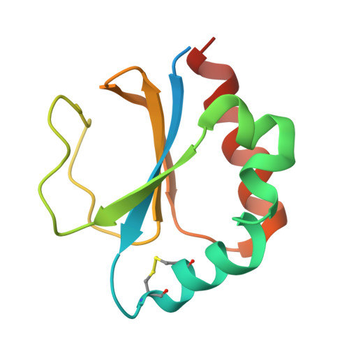
The structure of the periplasmic thiol-disulfide oxidoreductase SoxS from Paracoccus pantotrophus indicates a triple Trx/Grx/DsbC functionality in chemotrophic sulfur oxidation.

Carius, Y.Rother, D.Friedrich, C.G.Scheidig, A.J.

(2009) Acta Crystallogr D Biol Crystallogr 65: 229-240

  • DOI: https://doi.org/10.1107/S0907444908043023
  • Primary Citation Related Structures: 
    3D4T, 3DML

  • PubMed Abstract: 

    The periplasmic thiol-disulfide oxidoreductase SoxS is beneficial for the sulfur-oxidizing (Sox) phenotype of the facultative chemotrophic bacterium Paracoccus pantotrophus and is not part of the Sox enzyme system. SoxS combines features of thioredoxins, glutaredoxins and the thiol-disulfide oxidoreductases of the Dsb family in structure, target specificity and reaction. The structure of SoxS was solved in oxidized and reduced forms at 2.1 and 1.9 A resolution, respectively. SoxS revealed high structural homology to typical cytoplasmic bacterial thioredoxins. In contrast, SoxS contained the active-site motif Pro-Gly-Cys-Leu-Tyr-Cys that is not present in other thioredoxins. Interestingly, the sequence of this motif is closely related to the Pro-Gly-Cys-Pro-Tyr-Cys sequence of some glutaredoxins and to the Pro-Xaa-Cys-Xaa-Tyr-Cys sequences of some members of the DsbC and DsbG subfamilies of thiol-disulfide oxidoreductases. Furthermore, the proposed substrate of SoxS, the interprotein disulfide of SoxY, Cys110(Y)-Cys110(Y), is structurally similar to oxidized glutathione. However, SoxS is proposed to specifically reduce the interprotein disulfide between two SoxY subunits, releasing a heterodimeric SoxYZ as an active part of the sulfur-oxidation cycle.


  • Organizational Affiliation
    • Abteilung für Strukturbiologie, Zoologisches Institut, Christian-Albrechts-Universität zu Kiel, Kiel, Germany.

Macromolecule Content 

  • Total Structure Weight: 13.24 kDa 
  • Atom Count: 904 
  • Modeled Residue Count: 97 
  • Deposited Residue Count: 116 
  • Unique protein chains: 1

Macromolecules

Find similar proteins by:|  3D Structure
Entity ID: 1
MoleculeChains  Sequence LengthOrganismDetailsImage
Putative uncharacterized protein116Paracoccus denitrificansMutation(s): 0 
Gene Names: soxS
UniProt
Find proteins for Q8KM22 (Paracoccus denitrificans)
Explore Q8KM22 
Go to UniProtKB:  Q8KM22
Entity Groups
Sequence Clusters30% Identity50% Identity70% Identity90% Identity95% Identity100% Identity
UniProt GroupQ8KM22
Sequence Annotations
Expand
Reference Sequence

Experimental Data & Validation

Experimental Data

  • Method: X-RAY DIFFRACTION
  • Resolution: 2.05 Å
  • R-Value Free:  0.216 (Depositor), 0.217 (DCC) 
  • R-Value Work:  0.175 (Depositor), 0.173 (DCC) 
  • R-Value Observed: 0.177 (Depositor) 
Space Group: P 31 2 1
Unit Cell:
Length ( Å )Angle ( ˚ )
a = 81α = 90
b = 81β = 90
c = 33.1γ = 120
Software Package:
Software NamePurpose
REFMACrefinement
CrysalisProdata collection
CrysalisProdata reduction
SCALAdata scaling
MOLREPphasing

Structure Validation

View Full Validation Report


Entry History 

Deposition Data

Revision History  (Full details and data files)

  • Version 1.0: 2008-07-15
    Type: Initial release
  • Version 1.1: 2011-07-13
    Changes: Version format compliance
  • Version 1.2: 2017-10-25
    Changes: Refinement description
  • Version 1.3: 2024-04-03
    Changes: Data collection, Database references, Derived calculations, Refinement description
  • Version 1.4: 2025-08-06
    Changes: Database references, Structure summary