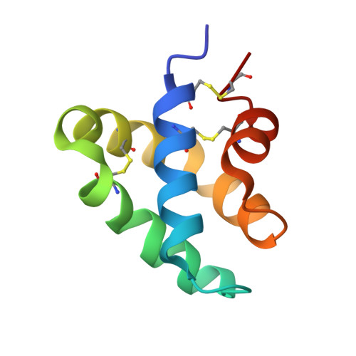
3BQQ

Crystal Structure of Human Saposin D (triclinic)


Experimental Data Snapshot

  • Method: X-RAY DIFFRACTION
  • Resolution: 2.00 Å
  • R-Value Free: 0.242 
  • R-Value Work: 0.195 
  • R-Value Observed: 0.198 

Starting Model: experimental
View more details

wwPDB Validation   3D Report Full Report


This is version 1.3 of the entry. See complete history


Literature

Structures of the human ceramide activator protein saposin D.

Popovic, K.Prive, G.G.

(2008) Acta Crystallogr D Biol Crystallogr 64: 589-594

  • DOI: https://doi.org/10.1107/S0907444908003120
  • Primary Citation of Related Structures:  
    3BQP, 3BQQ

  • PubMed Abstract: 

    Saposin D is a sphingolipid activator protein required for the lysosomal breakdown of ceramide to a fatty acid and sphingosine by acid ceramidase. The crystal structure of saposin D has been determined in two different crystal forms, resulting in a total of six crystallographically independent views of this small 80-amino-acid protein. All of the structures are highly similar and reveal the monomeric form of the saposin fold previously seen in the crystal structures of saposins A and C. Saposin D is slightly more compact than the related saposins A and C owing to a slight repositioning of the 'stem' and 'hairpin' regions of the protein.


  • Organizational Affiliation

    Department of Medical Biophysics, University of Toronto, Toronto, Ontario, Canada.


Macromolecules
Find similar proteins by:  (by identity cutoff)  |  3D Structure
Entity ID: 1
MoleculeChains Sequence LengthOrganismDetailsImage
PROACTIVATOR POLYPEPTIDEA [auth C],
B [auth A],
C [auth B],
D
80Homo sapiensMutation(s): 0 
Gene Names: PSAP
UniProt & NIH Common Fund Data Resources
Find proteins for P07602 (Homo sapiens)
Explore P07602 
Go to UniProtKB:  P07602
PHAROS:  P07602
GTEx:  ENSG00000197746 
Entity Groups  
Sequence Clusters30% Identity50% Identity70% Identity90% Identity95% Identity100% Identity
UniProt GroupP07602
Sequence Annotations
Expand
  • Reference Sequence
Experimental Data & Validation

Experimental Data

  • Method: X-RAY DIFFRACTION
  • Resolution: 2.00 Å
  • R-Value Free: 0.242 
  • R-Value Work: 0.195 
  • R-Value Observed: 0.198 
  • Space Group: P 1
Unit Cell:
Length ( Å )Angle ( ˚ )
a = 40.66α = 89.9
b = 41.12β = 81.2
c = 65.45γ = 71.4
Software Package:
Software NamePurpose
PHASERphasing
REFMACrefinement
PROTEUM PLUSdata collection
PROTEUM PLUSdata reduction
PROTEUM PLUSdata scaling

Structure Validation

View Full Validation Report


Entry History 

Deposition Data

Revision History  (Full details and data files)

  • Version 1.0: 2008-02-05
    Type: Initial release
  • Version 1.1: 2011-07-13
    Changes: Version format compliance
  • Version 1.2: 2024-04-03
    Changes: Data collection, Database references, Derived calculations, Refinement description
  • Version 1.3: 2024-11-06
    Changes: Structure summary