3BE6

Crystal structure of FitE (crystal form 2), a group III periplasmic siderophore binding protein


Experimental Data Snapshot

  • Method: X-RAY DIFFRACTION
  • Resolution: 1.82 Å
  • R-Value Free: 0.219 
  • R-Value Work: 0.188 
  • R-Value Observed: 0.190 

wwPDB Validation   3D Report Full Report


This is version 1.3 of the entry. See complete history


Literature

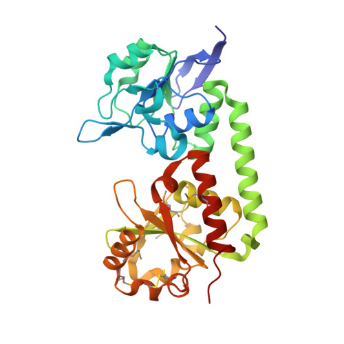
Trapping open and closed forms of FitE-A group III periplasmic binding protein.

Shi, R.Proteau, A.Wagner, J.Cui, Q.Purisima, E.O.Matte, A.Cygler, M.

(2008) Proteins 75: 598-609

  • DOI: https://doi.org/10.1002/prot.22272
  • Primary Citation of Related Structures:  
    3BE5, 3BE6

  • PubMed Abstract: 

    Periplasmic binding proteins (PBPs) are essential components of bacterial transport systems, necessary for bacterial growth and survival. The two-domain structures of PBPs are topologically classified into three groups based on the number of crossovers or hinges between the globular domains: group I PBPs have three connections, group II have two, and group III have only one. Although a large number of structures for group I or II PBPs are known, fewer group III PBPs have been structurally characterized. Group I and II PBPs exhibit significant domain motions during transition from the unbound to ligand-bound form, however, no large conformational changes have been observed to date in group III PBPs. We have solved the crystal structure of a periplasmic binding protein FitE, part of an iron transport system, fit, recently identified in a clinical E. coli isolate. The structure, determined at 1.8 A resolution, shows that FitE is a group III PBP containing a single alpha-helix bridging the two domains. Among the individual FitE molecules present in two crystal forms we observed three different conformations (open, closed, intermediate). Our crystallographic and molecular dynamics results strongly support the notion that group III PBPs also adopt the same Venus flytrap mechanism as do groups I and II PBPs. Unlike other group III PBPs, FitE forms dimers both in solution and in the crystals. The putative siderophore binding pocket is lined with arginine residues, suggesting an anionic nature of the iron-containing siderophore.


  • Organizational Affiliation

    Department of Biochemistry, McGill University, Montréal, Québec, Canada.


Macromolecules
Find similar proteins by:  (by identity cutoff)  |  3D Structure
Entity ID: 1
MoleculeChains Sequence LengthOrganismDetailsImage
Putative iron compound-binding protein of ABC transporter family
A, B, C, D
297N/AMutation(s): 0 
Gene Names: fiteECs3913Z4382fitE
UniProt
Find proteins for Q8XBR1 (Escherichia coli O157:H7)
Explore Q8XBR1 
Go to UniProtKB:  Q8XBR1
Entity Groups  
Sequence Clusters30% Identity50% Identity70% Identity90% Identity95% Identity100% Identity
UniProt GroupQ8XBR1
Sequence Annotations
Expand
  • Reference Sequence
Small Molecules
Ligands 3 Unique
IDChains Name / Formula / InChI Key2D Diagram3D Interactions
GOL
Query on GOL

Download Ideal Coordinates CCD File 
G [auth A],
J [auth C],
O [auth D]
GLYCEROL
C3 H8 O3
PEDCQBHIVMGVHV-UHFFFAOYSA-N
CL
Query on CL

Download Ideal Coordinates CCD File 
E [auth A]
H [auth C]
I [auth C]
K [auth D]
L [auth D]
E [auth A],
H [auth C],
I [auth C],
K [auth D],
L [auth D],
M [auth D],
N [auth D]
CHLORIDE ION
Cl
VEXZGXHMUGYJMC-UHFFFAOYSA-M
MG
Query on MG

Download Ideal Coordinates CCD File 
F [auth A]MAGNESIUM ION
Mg
JLVVSXFLKOJNIY-UHFFFAOYSA-N
Modified Residues  1 Unique
IDChains TypeFormula2D DiagramParent
MSE
Query on MSE
A, B, C, D
L-PEPTIDE LINKINGC5 H11 N O2 SeMET
Experimental Data & Validation

Experimental Data

  • Method: X-RAY DIFFRACTION
  • Resolution: 1.82 Å
  • R-Value Free: 0.219 
  • R-Value Work: 0.188 
  • R-Value Observed: 0.190 
  • Space Group: P 21 21 21
Unit Cell:
Length ( Å )Angle ( ˚ )
a = 50.815α = 90
b = 109.12β = 90
c = 221.992γ = 90
Software Package:
Software NamePurpose
REFMACrefinement
ADSCdata collection
HKL-2000data reduction
HKL-2000data scaling
MOLREPphasing

Structure Validation

View Full Validation Report


Entry History 

Deposition Data

Revision History  (Full details and data files)

  • Version 1.0: 2008-10-28
    Type: Initial release
  • Version 1.1: 2011-07-13
    Changes: Advisory, Version format compliance
  • Version 1.2: 2023-08-30
    Changes: Data collection, Database references, Derived calculations, Refinement description
  • Version 1.3: 2023-11-15
    Changes: Data collection