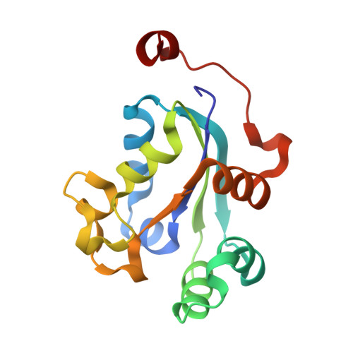
3BBF

Crystal structure of the NM23-H2 transcription factor complex with GDP


Experimental Data Snapshot

  • Method: X-RAY DIFFRACTION
  • Resolution: 1.70 Å
  • R-Value Free: 0.236 
  • R-Value Work: 0.196 
  • R-Value Observed: 0.198 

Starting Model: experimental
View more details

wwPDB Validation   3D Report Full Report


Ligand Structure Quality Assessment 


This is version 1.2 of the entry. See complete history


Literature

NM23-H2 may play an indirect role in transcriptional activation of c-myc gene expression but does not cleave the nuclease hypersensitive element III1.

Dexheimer, T.S.Carey, S.S.Zuohe, S.Gokhale, V.M.Hu, X.Murata, L.B.Maes, E.M.Weichsel, A.Sun, D.Meuillet, E.J.Montfort, W.R.Hurley, L.H.

(2009) Mol Cancer Ther 8: 1363-1377

  • DOI: https://doi.org/10.1158/1535-7163.MCT-08-1093
  • Primary Citation of Related Structures:  
    3BBB, 3BBC, 3BBF

  • PubMed Abstract: 

    The formation of G-quadruplex structures within the nuclease hypersensitive element (NHE) III(1) region of the c-myc promoter and the ability of these structures to repress c-myc transcription have been well established. However, just how these extremely stable DNA secondary structures are transformed to activate c-myc transcription is still unknown. NM23-H2/nucleoside diphosphate kinase B has been recognized as an activator of c-myc transcription via interactions with the NHE III(1) region of the c-myc gene promoter. Through the use of RNA interference, we confirmed the transcriptional regulatory role of NM23-H2. In addition, we find that further purification of NM23-H2 results in loss of the previously identified DNA strand cleavage activity, but retention of its DNA binding activity. NM23-H2 binds to both single-stranded guanine- and cytosine-rich strands of the c-myc NHE III(1) and, to a lesser extent, to a random single-stranded DNA template. However, it does not bind to or cleave the NHE III(1) in duplex form. Significantly, potassium ions and compounds that stabilize the G-quadruplex and i-motif structures have an inhibitory effect on NM23-H2 DNA-binding activity. Mutation of Arg(88) to Ala(88) (R88A) reduced both DNA and nucleotide binding but had minimal effect on the NM23-H2 crystal structure. On the basis of these data and molecular modeling studies, we have proposed a stepwise trapping-out of the NHE III(1) region in a single-stranded form, thus allowing single-stranded transcription factors to bind and activate c-myc transcription. Furthermore, this model provides a rationale for how the stabilization of the G-quadruplex or i-motif structures formed within the c-myc gene promoter region can inhibit NM23-H2 from activating c-myc gene expression.


  • Organizational Affiliation

    College of Pharmacy,University of Arizona, Tucson, Arizona 85721, USA.


Macromolecules
Find similar proteins by:  (by identity cutoff)  |  3D Structure
Entity ID: 1
MoleculeChains Sequence LengthOrganismDetailsImage
Nucleoside diphosphate kinase B
A, B, C, D, E
A, B, C, D, E, F
151Homo sapiensMutation(s): 0 
Gene Names: NME2NM23B
EC: 2.7.4.6
UniProt & NIH Common Fund Data Resources
Find proteins for P22392 (Homo sapiens)
Explore P22392 
Go to UniProtKB:  P22392
PHAROS:  P22392
GTEx:  ENSG00000243678 
Entity Groups  
Sequence Clusters30% Identity50% Identity70% Identity90% Identity95% Identity100% Identity
UniProt GroupP22392
Sequence Annotations
Expand
  • Reference Sequence
Small Molecules
Ligands 3 Unique
IDChains Name / Formula / InChI Key2D Diagram3D Interactions
GDP
Query on GDP

Download Ideal Coordinates CCD File 
G [auth A]
I [auth B]
L [auth C]
N [auth D]
P [auth E]
G [auth A],
I [auth B],
L [auth C],
N [auth D],
P [auth E],
R [auth F]
GUANOSINE-5'-DIPHOSPHATE
C10 H15 N5 O11 P2
QGWNDRXFNXRZMB-UUOKFMHZSA-N
DTT
Query on DTT

Download Ideal Coordinates CCD File 
K [auth B],
T [auth F]
2,3-DIHYDROXY-1,4-DITHIOBUTANE
C4 H10 O2 S2
VHJLVAABSRFDPM-IMJSIDKUSA-N
MG
Query on MG

Download Ideal Coordinates CCD File 
H [auth A]
J [auth B]
M [auth C]
O [auth D]
Q [auth E]
H [auth A],
J [auth B],
M [auth C],
O [auth D],
Q [auth E],
S [auth F]
MAGNESIUM ION
Mg
JLVVSXFLKOJNIY-UHFFFAOYSA-N
Binding Affinity Annotations 
IDSourceBinding Affinity
GDP PDBBind:  3BBF Kd: 5200 (nM) from 1 assay(s)
Experimental Data & Validation

Experimental Data

  • Method: X-RAY DIFFRACTION
  • Resolution: 1.70 Å
  • R-Value Free: 0.236 
  • R-Value Work: 0.196 
  • R-Value Observed: 0.198 
  • Space Group: P 21 21 21
Unit Cell:
Length ( Å )Angle ( ˚ )
a = 69.75α = 90
b = 86.78β = 90
c = 159.12γ = 90
Software Package:
Software NamePurpose
REFMACrefinement
XDISPLAYFdata collection
CrystalCleardata reduction
CrystalCleardata scaling
MOLREPphasing

Structure Validation

View Full Validation Report


Ligand Structure Quality Assessment 


Entry History 

Deposition Data

Revision History  (Full details and data files)

  • Version 1.0: 2008-09-23
    Type: Initial release
  • Version 1.1: 2011-07-13
    Changes: Version format compliance
  • Version 1.2: 2023-08-30
    Changes: Data collection, Database references, Derived calculations, Refinement description