Crystal structure of (Gly-Pro-Hyp)(9) : Implications for the collagen molecular model.
Okuyama, K., Miyama, K., Mizuno, K., Bachinger, H.P.(2012) Biopolymers 97: 607-616
- PubMed: 22605552 Search on PubMed
- DOI: https://doi.org/10.1002/bip.22048
- Primary Citation Related Structures:
3B0S - PubMed Abstract:
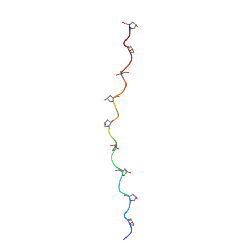
Collagens have long been believed to adopt a triple-stranded molecular structure with a 10/3 symmetry (ten triplet units in three turns) and an axial repeat of 29 Å. This belief even persisted after an alternative structure with a 7/2 symmetry (seven triplet units in two turns) with an axial repeat of 20 Å had been proposed. The uncertainty regarding the helical symmetry of collagens is attributed to inadequate X-ray fiber diffraction data. Therefore, for better understanding of the collagen helix, single-crystal analyses of peptides with simplified characteristic amino acid sequences and similar compositions to collagens have long been awaited. Here we report the crystal structure of (Gly-Pro-Hyp)(9) peptide at a resolution of 1.45 Å. The repeating unit of this peptide, Gly-Pro-Hyp, is the most typical sequence present in collagens, and it has been used as a basic repeating unit in fiber diffraction analyses of collagen. The (Gly-Pro-Hyp)(9) peptide adopts a triple-stranded structure with an average helical symmetry close to the ideal 7/2 helical model for collagen. This observation strongly suggests that the average molecular structure of collagen is not the accepted Rich and Crick 10/3 helical model but is a 7/2 helical conformation.
- Department of Macromolecular Science, Osaka University, Toyonaka, Osaka, Japan. okuyamak@chem.sci.osaka-u.ac.jp
Organizational Affiliation: