3AZ1 | pdb_00003az1

Crystal Structure Analysis of Vitamin D receptor


Experimental Data Snapshot

  • Method: X-RAY DIFFRACTION
  • Resolution: 1.50 Å
  • R-Value Free: 
    0.213 (Depositor) 
  • R-Value Work: 
    0.195 (Depositor), 0.209 (DCC) 
  • R-Value Observed: 
    0.195 (Depositor) 

wwPDB Validation 3D Report Full Report

Validation slider image for 3AZ1

Ligand Structure Quality Assessment 


This is version 1.2 of the entry. See complete history

Literature

Novel nonsecosteroidal vitamin D(3) carboxylic acid analogs for osteoporosis, and SAR analysis.

Kashiwagi, H.Ono, Y.Shimizu, K.Haneishi, T.Ito, S.Iijima, S.Kobayashi, T.Ichikawa, F.Harada, S.Sato, H.Sekiguchi, N.Ishigai, M.Takahashi, T.

(2011) Bioorg Med Chem 19: 4721-4729

  • DOI: https://doi.org/10.1016/j.bmc.2011.07.001
  • Primary Citation Related Structures: 
    3AZ1, 3AZ2, 3AZ3

  • PubMed Abstract: 

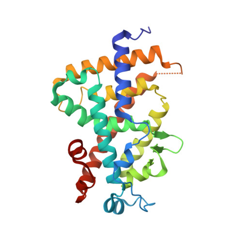
    Novel vitamin D(3) analogs with carboxylic acid were explored, focusing on a nonsecosteroidal analog, LG190178, with a bisphenyl skeleton. From X-ray analysis of these analogs with vitamin D receptor (VDR), the carboxyl groups had very unique hydrogen bonding interactions in VDR and mimicked 1α-hydroxy group and/or 3β-hydroxy group of 1α,25-dihydroxyvitamin D(3). A highly potent analog, 6a, with good in vitro activity and pharmacokinetic profiles was identified from an SAR study. Compound 6a showed significant prevention of bone loss in a rat osteoporosis model by oral administration.


  • Organizational Affiliation
    • Research Division, Chugai Pharmaceutical Co. Ltd, 1-135 Komakado, Gotemba, Shizuoka 412-8513, Japan. kashiwagihrt@chugai-pharm.co.jp

Macromolecule Content 

  • Total Structure Weight: 29.19 kDa 
  • Atom Count: 2,260 
  • Modeled Residue Count: 250 
  • Deposited Residue Count: 253 
  • Unique protein chains: 1

Macromolecules

Find similar proteins by:|  3D Structure
Entity ID: 1
MoleculeChains  Sequence LengthOrganismDetailsImage
Vitamin D3 receptor253Homo sapiensMutation(s): 0 
Gene Names: NR1I1VDR
UniProt & NIH Common Fund Data Resources
Find proteins for P11473 (Homo sapiens)
Explore P11473 
Go to UniProtKB:  P11473
PHAROS:  P11473
GTEx:  ENSG00000111424 
Entity Groups
Sequence Clusters30% Identity50% Identity70% Identity90% Identity95% Identity100% Identity
UniProt GroupP11473
Sequence Annotations
Expand
Reference Sequence

Small Molecules

Ligands 1 Unique
IDChains Name / Formula / InChI Key2D Diagram3D Interactions
DS2

Query on DS2


Download:Ideal Coordinates CCD File
B [auth A]{4-[3-(4-{[(2R)-2-hydroxy-3,3-dimethylbutyl]oxy}-3-methylphenyl)pentan-3-yl]-2-methylphenoxy}acetic acid
C27 H38 O5
PMGXXWMCGADHQY-DEOSSOPVSA-N

Experimental Data & Validation

Experimental Data

  • Method: X-RAY DIFFRACTION
  • Resolution: 1.50 Å
  • R-Value Free:  0.213 (Depositor) 
  • R-Value Work:  0.195 (Depositor), 0.209 (DCC) 
  • R-Value Observed: 0.195 (Depositor) 
Space Group: P 21 21 21
Unit Cell:
Length ( Å )Angle ( ˚ )
a = 44.613α = 90
b = 51.732β = 90
c = 131.579γ = 90
Software Package:
Software NamePurpose
MOSFLMdata reduction
SCALAdata scaling
BUSTER-TNTrefinement
PDB_EXTRACTdata extraction
BUSTERrefinement

Structure Validation

View Full Validation Report


Ligand Structure Quality Assessment 


Entry History 

Deposition Data

Revision History  (Full details and data files)

  • Version 1.0: 2011-11-02
    Type: Initial release
  • Version 1.1: 2017-08-23
    Changes: Refinement description, Source and taxonomy
  • Version 1.2: 2024-03-13
    Changes: Data collection, Database references, Derived calculations