3AYI

X-ray crystal structures of L-phenylalanine oxidase (deaminating and decaboxylating) from Pseudomonas sp. P501. Structures of the enzyme-ligand complex and catalytic mechanism


Experimental Data Snapshot

  • Method: X-RAY DIFFRACTION
  • Resolution: 1.25 Å
  • R-Value Free: 0.131 
  • R-Value Work: 0.104 
  • R-Value Observed: 0.105 

wwPDB Validation   3D Report Full Report


Ligand Structure Quality Assessment 


This is version 1.2 of the entry. See complete history


Literature

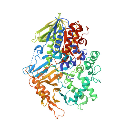
High resolution X-ray crystal structures of L-phenylalanine oxidase (deaminating and decarboxylating) from Pseudomonas sp. P-501. Structures of the enzyme-ligand complex and catalytic mechanism

Ida, K.Suguro, M.Suzuki, H.

(2011) J Biochem 150: 659-669

  • DOI: https://doi.org/10.1093/jb/mvr103
  • Primary Citation of Related Structures:  
    3AYI, 3AYJ, 3AYL

  • PubMed Abstract: 

    The mature form of L-Phe oxidase of Pseudomonas sp. P-501 (PAOpt) catalyzes the oxygenative decarboxylation of L-Phe and the oxidative deamination of L-Met, and is highly specific for L-Phe. The crystal structures of PAOpt individually complexed with L-Phe and L-Met and the properties of the active site mutants were investigated to clarify the structural basis of the substrate and reaction specificities of the enzyme. The benzene ring of L-Phe is packed in six hydrophobic amino acid side chains versus the two hydrophobic side chains of L-amino acid oxidase (LAO, pdb code: 2jb2); the distance between the substrate Cα atom and water is shorter in the PAOpt-L-Met complex than in the PAOpt-L-Phe complex; and the mutation of substrate carboxylate-binding residues (Arg143 and Tyr536) causes the enzyme to oxidize L-Phe and decreases the charge-transfer band with L-Phe. These results suggest that (i) the higher substrate specificity of PAOpt relative to LAO is derived from the compact hydrophobic nature of the PAOpt active site and (ii) the reactivity of the PAOpt charge-transfer complex with water or oxygen determines whether the enzyme catalyzes oxidation or oxygenation, respectively.


  • Organizational Affiliation

    Department of Biosciences, School of Science, Kitasato University, Kitasato 1-15-1, Sagamihara, Kanagawa 252-0329, Japan.


Macromolecules
Find similar proteins by:  (by identity cutoff)  |  3D Structure
Entity ID: 1
MoleculeChains Sequence LengthOrganismDetailsImage
Pro-enzyme of L-phenylalanine oxidase
A, B
721Pseudomonas sp. P-501Mutation(s): 0 
Gene Names: propao
EC: 1.13.12.9
UniProt
Find proteins for Q5W9R9 (Pseudomonas sp)
Explore Q5W9R9 
Go to UniProtKB:  Q5W9R9
Entity Groups  
Sequence Clusters30% Identity50% Identity70% Identity90% Identity95% Identity100% Identity
UniProt GroupQ5W9R9
Sequence Annotations
Expand
  • Reference Sequence
Small Molecules
Ligands 4 Unique
IDChains Name / Formula / InChI Key2D Diagram3D Interactions
FAD
Query on FAD

Download Ideal Coordinates CCD File 
D [auth A],
H [auth B]
FLAVIN-ADENINE DINUCLEOTIDE
C27 H33 N9 O15 P2
VWWQXMAJTJZDQX-UYBVJOGSSA-N
HCI
Query on HCI

Download Ideal Coordinates CCD File 
E [auth A],
I [auth B]
HYDROCINNAMIC ACID
C9 H10 O2
XMIIGOLPHOKFCH-UHFFFAOYSA-N
SO4
Query on SO4

Download Ideal Coordinates CCD File 
C [auth A],
G [auth B]
SULFATE ION
O4 S
QAOWNCQODCNURD-UHFFFAOYSA-L
GOL
Query on GOL

Download Ideal Coordinates CCD File 
F [auth A],
J [auth B]
GLYCEROL
C3 H8 O3
PEDCQBHIVMGVHV-UHFFFAOYSA-N
Binding Affinity Annotations 
IDSourceBinding Affinity
HCI Binding MOAD:  3AYI Ki: 1.39e+4 (nM) from 1 assay(s)
Experimental Data & Validation

Experimental Data

  • Method: X-RAY DIFFRACTION
  • Resolution: 1.25 Å
  • R-Value Free: 0.131 
  • R-Value Work: 0.104 
  • R-Value Observed: 0.105 
  • Space Group: P 21 21 21
Unit Cell:
Length ( Å )Angle ( ˚ )
a = 101.29α = 90
b = 112.694β = 90
c = 136.542γ = 90
Software Package:
Software NamePurpose
REFMACrefinement
MOSFLMdata reduction
SCALAdata scaling

Structure Validation

View Full Validation Report


Ligand Structure Quality Assessment 


Entry History 

Deposition Data

Revision History  (Full details and data files)

  • Version 1.0: 2011-08-31
    Type: Initial release
  • Version 1.1: 2013-06-19
    Changes: Database references
  • Version 1.2: 2023-11-01
    Changes: Data collection, Database references, Derived calculations, Refinement description