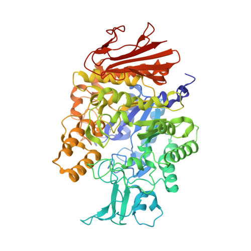
Steric hindrance by 2 amino acid residues determines the substrate specificity of isomaltase from Saccharomyces cerevisiae
Yamamoto, K., Miyake, H., Kusunoki, M., Osaki, S.(2011) J Biosci Bioeng 112: 545-550
- PubMed: 21925939 Search on PubMed
- DOI: https://doi.org/10.1016/j.jbiosc.2011.08.016
- Primary Citation Related Structures:
3AXH, 3AXI - PubMed Abstract:
The structures of the E277A isomaltase mutant from Saccharomyces cerevisiae in complex with isomaltose or maltose were determined at resolutions of 1.80 and 1.40Å, respectively. The root mean square deviations between the corresponding main-chain atoms of free isomaltase and the E277Α-isomaltose complex structures and those of free isomaltase and the E277A-maltose complex structures were found to be 0.131Å and 0.083Å, respectively. Thus, the amino acid substitution and ligand binding do not affect the overall structure of isomaltase. In the E277A-isomaltose structure, the bound isomaltose was readily identified by electron densities in the active site pocket; however, the reducing end of maltose was not observed in the E277A-maltose structure. The superposition of maltose onto the E277A-maltose structure revealed that the reducing end of maltose cannot bind to the subsite +1 due to the steric hindrance from Val216 and Gln279. The amino acid sequence comparisons with α-glucosidases showed that a bulky hydrophobic amino acid residue is conserved at the position of Val216 in α-1,6-glucosidic linkage hydrolyzing enzymes. Similarly, a bulky amino acid residue is conserved at the position of Gln279 in α-1,6-glucosidic linkage-only hydrolyzing α-glucosidases. Ala, Gly, or Asn residues were located at the position of α-1,4-glucosidic linkage hydrolyzing α-glucosidases. Two isomaltase mutant enzymes - V216T and Q279A - hydrolyzed maltose. Thus, the amino acid residues at these positions may be largely responsible for determining the substrate specificity of α-glucosidases.
- School of Medicine, Nara Medical University, 840 Shijo, Kashihara, Nara 634-8521, Japan. kama@naramed-u.ac.jp
Organizational Affiliation: