3ARU | pdb_00003aru

Crystal Structure Analysis of Chitinase A from Vibrio harveyi with novel inhibitors - W275G mutant complex structure with PENTOXIFYLLINE


Experimental Data Snapshot

  • Method: X-RAY DIFFRACTION
  • Resolution: 1.90 Å
  • R-Value Free: 
    0.195 (Depositor), 0.195 (DCC) 
  • R-Value Work: 
    0.143 (Depositor), 0.143 (DCC) 
  • R-Value Observed: 
    0.146 (Depositor) 

Starting Model: experimental
View more details

wwPDB Validation 3D Report Full Report

Validation slider image for 3ARU

Ligand Structure Quality Assessment 


This is version 1.4 of the entry. See complete history

Literature

Potent family-18 chitinase inhibitors: x-ray structures, affinities, and binding mechanisms

Pantoom, S.Vetter, I.R.Prinz, H.Suginta, W.

(2011) J Biological Chem 286: 24312-24323

  • DOI: https://doi.org/10.1074/jbc.M110.183376
  • Primary Citation Related Structures: 
    3ARO, 3ARP, 3ARQ, 3ARR, 3ARS, 3ART, 3ARU, 3ARV, 3ARW, 3ARX, 3ARY, 3ARZ, 3AS0, 3AS1, 3AS2, 3AS3

  • PubMed Abstract: 

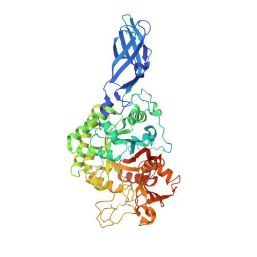
    Six novel inhibitors of Vibrio harveyi chitinase A (VhChiA), a family-18 chitinase homolog, were identified by in vitro screening of a library of pharmacologically active compounds. Unlike the previously identified inhibitors that mimicked the reaction intermediates, crystallographic evidence from 14 VhChiA-inhibitor complexes showed that all of the inhibitor molecules occupied the outer part of the substrate-binding cleft at two hydrophobic areas. The interactions at the aglycone location are well defined and tightly associated with Trp-397 and Trp-275, whereas the interactions at the glycone location are patchy, indicating lower affinity and a loose interaction with two consensus residues, Trp-168 and Val-205. When Trp-275 was substituted with glycine (W275G), the binding affinity toward all of the inhibitors dramatically decreased, and in most structures two inhibitor molecules were found to stack against Trp-397 at the aglycone site. Such results indicate that hydrophobic interactions are important for binding of the newly identified inhibitors by the chitinase. X-ray data and isothermal microcalorimetry showed that the inhibitors occupied the active site of VhChiA in three different binding modes, including single-site binding, independent two-site binding, and sequential two-site binding. The inhibitory effect of dequalinium in the low nanomolar range makes this compound an extremely attractive lead compound for plausible development of therapeutics against human diseases involving chitinase-mediated pathologies.


  • Organizational Affiliation
    • Biochemistry-Electrochemistry Research Unit, School of Chemistry, Institute of Science, Suranaree University of Technology, Nakhon Ratchasima 30000, Thailand.

Macromolecule Content 

  • Total Structure Weight: 64.55 kDa 
  • Atom Count: 5,129 
  • Modeled Residue Count: 570 
  • Deposited Residue Count: 584 
  • Unique protein chains: 1

Macromolecules

Find similar proteins by:|  3D Structure
Entity ID: 1
MoleculeChains  Sequence LengthOrganismDetailsImage
Chitinase A584Vibrio harveyiMutation(s): 1 
Gene Names: CHIA
EC: 3.2.1.14
UniProt
Find proteins for Q9AMP1 (Vibrio harveyi)
Explore Q9AMP1 
Go to UniProtKB:  Q9AMP1
Entity Groups
Sequence Clusters30% Identity50% Identity70% Identity90% Identity95% Identity100% Identity
UniProt GroupQ9AMP1
Sequence Annotations
Expand
Reference Sequence

Experimental Data & Validation

Experimental Data

  • Method: X-RAY DIFFRACTION
  • Resolution: 1.90 Å
  • R-Value Free:  0.195 (Depositor), 0.195 (DCC) 
  • R-Value Work:  0.143 (Depositor), 0.143 (DCC) 
  • R-Value Observed: 0.146 (Depositor) 
Space Group: P 21 21 21
Unit Cell:
Length ( Å )Angle ( ˚ )
a = 66.67α = 90
b = 83.93β = 90
c = 102.42γ = 90
Software Package:
Software NamePurpose
XSCALEdata scaling
REFMACrefinement
PDB_EXTRACTdata extraction

Structure Validation

View Full Validation Report


Ligand Structure Quality Assessment 


Entry History 

Deposition Data

Revision History  (Full details and data files)

  • Version 1.0: 2011-04-20
    Type: Initial release
  • Version 1.1: 2011-07-13
    Changes: Version format compliance
  • Version 1.2: 2014-01-29
    Changes: Database references
  • Version 1.3: 2023-11-01
    Changes: Data collection, Database references, Derived calculations, Refinement description
  • Version 1.4: 2024-11-13
    Changes: Structure summary