3AP7

Crystal structure of the galectin-8 N-terminal carbohydrate recognition domain in complex with lactose sialic acid


Experimental Data Snapshot

  • Method: X-RAY DIFFRACTION
  • Resolution: 1.53 Å
  • R-Value Free: 0.188 
  • R-Value Work: 0.164 
  • R-Value Observed: 0.165 

wwPDB Validation   3D Report Full Report


This is version 2.1 of the entry. See complete history


Literature

Galectin-8-N-domain recognition mechanism for sialylated and sulfated glycans

Ideo, H.Matsuzaka, T.Nonaka, T.Seko, A.Yamashita, K.

(2011) J Biol Chem 286: 11346-11355

  • DOI: https://doi.org/10.1074/jbc.M110.195925
  • Primary Citation of Related Structures:  
    3AP4, 3AP5, 3AP6, 3AP7, 3AP9

  • PubMed Abstract: 

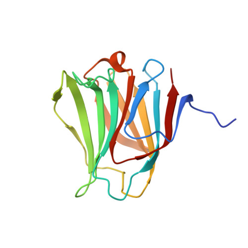
    Galectin-8 has much higher affinity for 3'-O-sulfated or 3'-O-sialylated glycoconjugates and a Lewis X-containing glycan than for oligosaccharides terminating in Galβ1→3/4GlcNAc, and this specificity is mainly attributed to the N-terminal carbohydrate recognition domain (N-domain, CRD) (Ideo, H., Seko, A., Ishizuka, I., and Yamashita, K. (2003) Glycobiology 13, 713-723). In this study, we elucidated the crystal structures of the human galectin-8-N-domain (-8N) in the absence or presence of 4 ligands. The apo molecule forms a dimer, which is different from the canonical 2-fold symmetric dimer observed for galectin-1 and -2. In a galectin-8N-lactose complex, the lactose-recognizing amino acids are highly conserved among the galectins. However, Arg(45), Gln(47), Arg(59), and the long loop region between the S3 and S4 β-strands are unique to galectin-8N. These amino acids directly or indirectly interact with the sulfate or sialic acid moieties of 3'-sialyl- and 3'-sulfolactose complexed with galectin-8N. Furthermore, in the LNF-III-galectin-8N complex, van der Waals interactions occur between the α1-3-branched fucose and galactose and between galactose and Tyr(141), and these interactions increase the affinity toward galectin-8N. Based on the findings of these x-ray crystallographic analyses, a mutagenesis study using surface plasmon resonance showed that Arg(45), Gln(47), and Arg(59) of galectin-8N are indispensable and coordinately contribute to the strong binding of galectins-8N to sialylated and sulfated oligosaccharides. Arg(59) is the most critical amino acid for binding in the S3-S4 loop region.


  • Organizational Affiliation

    Innovative Research Initiatives, Tokyo Institute of Technology, Yokohama, Japan.


Macromolecules
Find similar proteins by:  (by identity cutoff)  |  3D Structure
Entity ID: 1
MoleculeChains Sequence LengthOrganismDetailsImage
Galectin-8154Homo sapiensMutation(s): 0 
Gene Names: LGALS8
UniProt & NIH Common Fund Data Resources
Find proteins for O00214 (Homo sapiens)
Explore O00214 
Go to UniProtKB:  O00214
PHAROS:  O00214
GTEx:  ENSG00000116977 
Entity Groups  
Sequence Clusters30% Identity50% Identity70% Identity90% Identity95% Identity100% Identity
UniProt GroupO00214
Sequence Annotations
Expand
  • Reference Sequence
Oligosaccharides

Help

Entity ID: 2
MoleculeChains Length2D Diagram Glycosylation3D Interactions
N-acetyl-alpha-neuraminic acid-(2-3)-beta-D-galactopyranose-(1-4)-beta-D-glucopyranose
B
3N/A
Glycosylation Resources
GlyTouCan:  G91237TK
GlyCosmos:  G91237TK
GlyGen:  G91237TK
Small Molecules
Ligands 1 Unique
IDChains Name / Formula / InChI Key2D Diagram3D Interactions
CL
Query on CL

Download Ideal Coordinates CCD File 
C [auth A]CHLORIDE ION
Cl
VEXZGXHMUGYJMC-UHFFFAOYSA-M
Experimental Data & Validation

Experimental Data

  • Method: X-RAY DIFFRACTION
  • Resolution: 1.53 Å
  • R-Value Free: 0.188 
  • R-Value Work: 0.164 
  • R-Value Observed: 0.165 
  • Space Group: P 21 21 21
Unit Cell:
Length ( Å )Angle ( ˚ )
a = 46.36α = 90
b = 49.917β = 90
c = 70.289γ = 90
Software Package:
Software NamePurpose
ADSCdata collection
MOLREPphasing
REFMACrefinement
MOSFLMdata reduction
SCALAdata scaling

Structure Validation

View Full Validation Report


Entry History 

Deposition Data

Revision History  (Full details and data files)

  • Version 1.0: 2011-01-26
    Type: Initial release
  • Version 1.1: 2011-07-13
    Changes: Version format compliance
  • Version 1.2: 2014-01-15
    Changes: Database references
  • Version 2.0: 2020-07-29
    Type: Remediation
    Reason: Carbohydrate remediation
    Changes: Atomic model, Data collection, Derived calculations, Non-polymer description, Structure summary
  • Version 2.1: 2023-11-01
    Changes: Data collection, Database references, Derived calculations, Refinement description, Structure summary