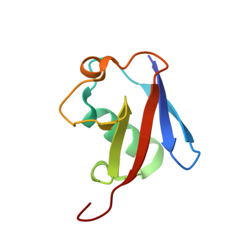
Crystal structure of cyclic Lys48-linked tetraubiquitin
Satoh, T., Sakata, E., Yamamoto, S., Yamaguchi, Y., Sumiyoshi, A., Wakatsuki, S., Kato, K.(2010) Biochem Biophys Res Commun
- PubMed: 20728431 Search on PubMed
- DOI: https://doi.org/10.1016/j.bbrc.2010.08.057
- Primary Citation Related Structures:
3ALB - PubMed Abstract:
Lys48-linked polyubiquitin chains serve as a signal for protein degradation by 26S proteasomes through its Ile44 hydrophobic patches interactions. The individual ubiquitin units of each chain are conjugated through an isopeptide bond between Lys48 and the C-terminal Gly76 of the preceding units. The conformation of Lys48-linked tetraubiquitin has been shown to change dynamically depending on solution pH. Here we enzymatically synthesized a wild-type Lys48-linked tetraubiquitin for structural study. In the synthesis, cyclic and non-cyclic species were obtained as major and minor fractions, respectively. This enabled us to solve the crystal structure of tetraubiquitin exclusively with native Lys48-linkages at 1.85A resolution in low pH 4.6. The crystallographic data clearly showed that the C-terminus of the first ubiquitin is conjugated to the Lys48 residue of the fourth ubiquitin. The overall structure is quite similar to the closed form of engineered tetraubiquitin at near-neutral pH 6.7, previously reported, in which the Ile44 hydrophobic patches face each other. The structure of the second and the third ubiquitin units [Ub(2)-Ub(3)] connected through a native isopeptide bond is significantly different from the conformations of the corresponding linkage of the engineered tetraubiquitins, whereas the structures of Ub(1)-Ub(2) and Ub(3)-Ub(4) isopeptide bonds are almost identical to those of the previously reported structures. From these observations, we suggest that the flexible nature of the isopeptide linkage thus observed contributes to the structural arrangements of ubiquitin chains exemplified by the pH-dependent closed-to-open conformational transition of tetraubiquitin.
- Structural Biology Research Center, Photon Factory, Institute of Materials Structure Science, High Energy Accelerator Research Organization (KEK), Tsukuba, Ibaraki 305-0801, Japan.
Organizational Affiliation: