3A5T | pdb_00003a5t

Crystal structure of MafG-DNA complex


Experimental Data Snapshot

  • Method: X-RAY DIFFRACTION
  • Resolution: 2.80 Å
  • R-Value Free: 
    0.294 (Depositor), 0.293 (DCC) 
  • R-Value Work: 
    0.261 (Depositor), 0.260 (DCC) 

wwPDB Validation 3D Report Full Report

Validation slider image for 3A5T

This is version 1.3 of the entry. See complete history

Literature

Structural Basis of Alternative DNA Recognition by Maf Transcription Factors

Kurokawa, H.Motohashi, H.Sueno, S.Kimura, M.Takagawa, H.Kanno, Y.Yamamoto, M.Tanaka, T.

(2009) Mol Cell Biol 29: 6232-6244

  • DOI: https://doi.org/10.1128/MCB.00708-09
  • Primary Citation Related Structures: 
    3A5T

  • PubMed Abstract: 

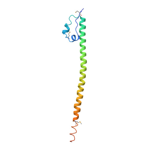
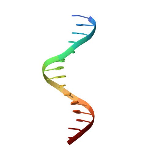
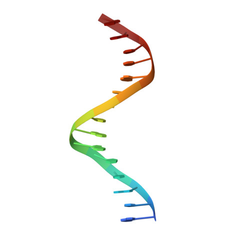
    Maf transcription factors constitute a family of the basic region-leucine zipper (bZip) factors and recognize unusually long DNA motifs (13 or 14 bp), termed the Maf recognition element (MARE). The MARE harbors extended GC sequences on each side of its core motif, which is similar to TRE or CRE (7 or 8 bp) recognized by the AP1 and CREB/ATF families, respectively. To ascertain the structural basis governing the acquirement of such unique DNA recognition, we determined the crystal structure of the MafG-DNA complex. Each MafG monomer consists of three helices in which the carboxyl-terminal long helix organizes one DNA-contacting element and one coiled-coil dimer formation element. To our surprise, two well-conserved residues, Arg57 and Asn61 in the basic region, play critical roles in Maf-specific DNA recognition. These two residues show unique side-chain orientations and interact directly with the extended GC bases. Maf-specific residues in the amino-terminal and basic regions appear to indirectly stabilize MARE recognition through DNA backbone phosphate interactions. This study revealed an alternative DNA recognition mechanism of the bZip factors that bestows specific target gene profiles upon Maf homodimers or Maf-containing heterodimers.


  • Organizational Affiliation
    • Institute of Multidisciplinary Research for Advanced Materials, Tohoku University, Sendai 980-8577, Japan. hkuro@m.tains.tohoku.ac.jp

Macromolecule Content 

  • Total Structure Weight: 34.25 kDa 
  • Atom Count: 2,129 
  • Modeled Residue Count: 216 
  • Deposited Residue Count: 244 
  • Unique protein chains: 1
  • Unique nucleic acid chains: 2

Macromolecules


Find similar proteins by:|  3D Structure
Entity ID: 1
MoleculeChains  Sequence LengthOrganismDetailsImage
Transcription factor MafG
A, B
107Mus musculusMutation(s): 0 
Gene Names: Mafg
UniProt
Find proteins for O54790 (Mus musculus)
Explore O54790 
Go to UniProtKB:  O54790
Entity Groups
Sequence Clusters30% Identity50% Identity70% Identity90% Identity95% Identity100% Identity
UniProt GroupO54790
Sequence Annotations
Expand
Reference Sequence
Find similar nucleic acids by:  Sequence
Entity ID: 2
MoleculeChains LengthOrganismImage
5'-D(*CP*TP*GP*AP*TP*GP*AP*GP*TP*CP*AP*GP*CP*AP*C)-3'15synthetic construct
Sequence Annotations
Expand
Reference Sequence
Find similar nucleic acids by:  Sequence
Entity ID: 3
MoleculeChains LengthOrganismImage
5'-D(*GP*TP*GP*CP*TP*GP*AP*CP*TP*CP*AP*TP*CP*AP*G)-3'15synthetic construct
Sequence Annotations
Expand
Reference Sequence

Small Molecules

Ligands 1 Unique
IDChains Name / Formula / InChI Key2D Diagram3D Interactions
MG

Query on MG


Download:Ideal Coordinates CCD File
E [auth C]MAGNESIUM ION
Mg
JLVVSXFLKOJNIY-UHFFFAOYSA-N
Modified Residues  1 Unique
IDChains TypeFormula2D DiagramParent
MSE
Query on MSE
A, B
L-PEPTIDE LINKINGC5 H11 N O2 SeMET

Experimental Data & Validation

Experimental Data

  • Method: X-RAY DIFFRACTION
  • Resolution: 2.80 Å
  • R-Value Free:  0.294 (Depositor), 0.293 (DCC) 
  • R-Value Work:  0.261 (Depositor), 0.260 (DCC) 
Space Group: C 2 2 21
Unit Cell:
Length ( Å )Angle ( ˚ )
a = 40.364α = 90
b = 185.243β = 90
c = 230.263γ = 90
Software Package:
Software NamePurpose
ADSCdata collection
SHELXDEphasing
CNSrefinement
HKL-2000data reduction
HKL-2000data scaling

Structure Validation

View Full Validation Report


Entry History 

Deposition Data

Revision History  (Full details and data files)

  • Version 1.0: 2009-10-13
    Type: Initial release
  • Version 1.1: 2011-07-13
    Changes: Version format compliance
  • Version 1.2: 2023-06-14
    Changes: Database references, Derived calculations, Source and taxonomy
  • Version 1.3: 2024-10-23
    Changes: Data collection, Structure summary