3A4S

The crystal structure of the SLD2:Ubc9 complex


Experimental Data Snapshot

  • Method: X-RAY DIFFRACTION
  • Resolution: 2.70 Å
  • R-Value Free: 0.269 
  • R-Value Work: 0.222 
  • R-Value Observed: 0.227 

wwPDB Validation   3D Report Full Report


This is version 1.2 of the entry. See complete history


Literature

Structural basis for regulation of poly-SUMO chain by a SUMO-like domain of Nip45

Sekiyama, N.Arita, K.Ikeda, Y.Hashiguchi, K.Ariyoshi, M.Tochio, H.Saitoh, H.Shirakawa, M.

(2009) Proteins 78: 1491-1502

  • DOI: https://doi.org/10.1002/prot.22667
  • Primary Citation of Related Structures:  
    3A4R, 3A4S

  • PubMed Abstract: 

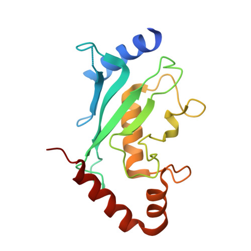
    Post-translational modification by small ubiquitin-like modifier (SUMO) provides an important regulatory mechanism in diverse cellular processes. Modification of SUMO has been shown to target proteins involved in systems ranging from DNA repair pathways to the ubiquitin-proteasome degradation system by the action of SUMO-targeted ubiquitin ligases (STUbLs). STUbLs recognize target proteins modified with a poly-SUMO chain through their SUMO-interacting motifs (SIMs). STUbLs are also associated with RENi family proteins, which commonly have two SUMO-like domains (SLD1 and SLD2) at their C terminus. We have determined the crystal structures of SLD2 of mouse RENi protein, Nip45, in a free form and in complex with a mouse E2 sumoylation enzyme, Ubc9. While Nip45 SLD2 shares a beta-grasp fold with SUMO, the SIM interaction surface conserved in SUMO paralogues does not exist in SLD2. Biochemical data indicates that neither tandem SLDs or SLD2 of Nip45 bind to either tandem SIMs from either mouse STUbL, RNF4 or to those from SUMO-binding proteins, whose interactions with SUMO have been well characterized. On the other hand, Nip45 SLD2 binds to Ubc9 in an almost identical manner to that of SUMO and thereby inhibits elongation of poly-SUMO chains. This finding highlights a possible role of the RENi proteins in the modulation of Ubc9-mediated poly-SUMO formation.


  • Organizational Affiliation

    Kyoto University, Japan.


Macromolecules
Find similar proteins by:  (by identity cutoff)  |  3D Structure
Entity ID: 1
MoleculeChains Sequence LengthOrganismDetailsImage
SUMO-conjugating enzyme UBC9
A, B
163Homo sapiensMutation(s): 0 
Gene Names: Ubc9
EC: 6.3.2
UniProt & NIH Common Fund Data Resources
Find proteins for P63279 (Homo sapiens)
Explore P63279 
Go to UniProtKB:  P63279
PHAROS:  P63279
GTEx:  ENSG00000103275 
Entity Groups  
Sequence Clusters30% Identity50% Identity70% Identity90% Identity95% Identity100% Identity
UniProt GroupP63279
Sequence Annotations
Expand
  • Reference Sequence
Find similar proteins by:  (by identity cutoff)  |  3D Structure
Entity ID: 2
MoleculeChains Sequence LengthOrganismDetailsImage
NFATC2-interacting protein
C, D
79Mus musculusMutation(s): 0 
Gene Names: Nip45
UniProt & NIH Common Fund Data Resources
Find proteins for O09130 (Mus musculus)
Explore O09130 
Go to UniProtKB:  O09130
IMPC:  MGI:1329015
Entity Groups  
Sequence Clusters30% Identity50% Identity70% Identity90% Identity95% Identity100% Identity
UniProt GroupO09130
Sequence Annotations
Expand
  • Reference Sequence
Experimental Data & Validation

Experimental Data

  • Method: X-RAY DIFFRACTION
  • Resolution: 2.70 Å
  • R-Value Free: 0.269 
  • R-Value Work: 0.222 
  • R-Value Observed: 0.227 
  • Space Group: P 1
Unit Cell:
Length ( Å )Angle ( ˚ )
a = 29.25α = 103.2
b = 49.42β = 92.1
c = 90.295γ = 101.13
Software Package:
Software NamePurpose
HKL-2000data collection
PHASERphasing
PHENIXrefinement
HKL-2000data reduction
HKL-2000data scaling

Structure Validation

View Full Validation Report


Entry History 

Deposition Data

Revision History  (Full details and data files)

  • Version 1.0: 2010-02-02
    Type: Initial release
  • Version 1.1: 2011-07-13
    Changes: Version format compliance
  • Version 1.2: 2023-11-01
    Changes: Data collection, Database references, Refinement description