2ZNX

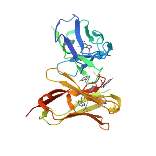
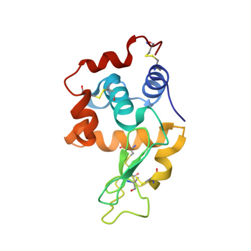
5-Fluorotryptophan Incorporated ScFv10 Complexed to Hen Egg Lysozyme


Experimental Data Snapshot

  • Method: X-RAY DIFFRACTION
  • Resolution: 2.30 Å
  • R-Value Free: 0.232 
  • R-Value Work: 0.193 
  • R-Value Observed: 0.195 

Starting Model: experimental
View more details

wwPDB Validation   3D Report Full Report


This is version 1.5 of the entry. See complete history


Literature

Specific fluorine labeling of the HyHEL10 antibody affects antigen binding and dynamics

Acchione, M.Lee, Y.C.DeSantis, M.E.Lipschultz, C.A.Wlodawer, A.Li, M.Shanmuganathan, A.Walter, R.L.Smith-Gill, S.Barchi, J.J.

(2012) Biochemistry 51: 6017-6027

  • DOI: https://doi.org/10.1021/bi300455t
  • Primary Citation of Related Structures:  
    2ZNW, 2ZNX

  • PubMed Abstract: 

    To more fully understand the molecular mechanisms responsible for variations in binding affinity with antibody maturation, we explored the use of site specific fluorine labeling and (19)F nuclear magnetic resonance (NMR). Several single-chain (scFv) antibodies, derived from an affinity-matured series of anti-hen egg white lysozyme (HEL) mouse IgG1, were constructed with either complete or individual replacement of tryptophan residues with 5-fluorotryptophan ((5F)W). An array of biophysical techniques was used to gain insight into the impact of fluorine substitution on the overall protein structure and antigen binding. SPR measurements indicated that (5F)W incorporation lowered binding affinity for the HEL antigen. The degree of analogue impact was residue-dependent, and the greatest decrease in affinity was observed when (5F)W was substituted for residues near the binding interface. In contrast, corresponding crystal structures in complex with HEL were essentially indistinguishable from the unsubstituted antibody. (19)F NMR analysis showed severe overlap of signals in the free fluorinated protein that was resolved upon binding to antigen, suggesting very distinct chemical environments for each (5F)W in the complex. Preliminary relaxation analysis suggested the presence of chemical exchange in the antibody-antigen complex that could not be observed by X-ray crystallography. These data demonstrate that fluorine NMR can be an extremely useful tool for discerning structural changes in scFv antibody-antigen complexes with altered function that may not be discernible by other biophysical techniques.


  • Organizational Affiliation

    Structural Biophysics Laboratory, Center for Cancer Research, Frederick National Laboratory for Cancer Research, Frederick, MD 21702, USA.


Macromolecules
Find similar proteins by:  (by identity cutoff)  |  3D Structure
Entity ID: 1
MoleculeChains Sequence LengthOrganismDetailsImage
ScFvA,
C [auth B]
242Mus musculussynthetic constructMutation(s): 0 
UniProt
Find proteins for Q65ZI1 (Mus musculus)
Explore Q65ZI1 
Go to UniProtKB:  Q65ZI1
Entity Groups  
Sequence Clusters30% Identity50% Identity70% Identity90% Identity95% Identity100% Identity
UniProt GroupQ65ZI1
Sequence Annotations
Expand
  • Reference Sequence
Find similar proteins by:  (by identity cutoff)  |  3D Structure
Entity ID: 2
MoleculeChains Sequence LengthOrganismDetailsImage
Lysozyme CB [auth Y],
D [auth Z]
129Gallus gallusMutation(s): 0 
EC: 3.2.1.17
UniProt
Find proteins for P00698 (Gallus gallus)
Explore P00698 
Go to UniProtKB:  P00698
Entity Groups  
Sequence Clusters30% Identity50% Identity70% Identity90% Identity95% Identity100% Identity
UniProt GroupP00698
Sequence Annotations
Expand
  • Reference Sequence
Small Molecules
Ligands 1 Unique
IDChains Name / Formula / InChI Key2D Diagram3D Interactions
1PG
Query on 1PG

Download Ideal Coordinates CCD File 
E [auth A],
F [auth B]
2-(2-{2-[2-(2-METHOXY-ETHOXY)-ETHOXY]-ETHOXY}-ETHOXY)-ETHANOL
C11 H24 O6
SLNYBUIEAMRFSZ-UHFFFAOYSA-N
Modified Residues  1 Unique
IDChains TypeFormula2D DiagramParent
FTR
Query on FTR
A,
C [auth B]
L-PEPTIDE LINKINGC11 H11 F N2 O2TRP
Experimental Data & Validation

Experimental Data

  • Method: X-RAY DIFFRACTION
  • Resolution: 2.30 Å
  • R-Value Free: 0.232 
  • R-Value Work: 0.193 
  • R-Value Observed: 0.195 
  • Space Group: P 41 21 2
Unit Cell:
Length ( Å )Angle ( ˚ )
a = 149.24α = 90
b = 149.24β = 90
c = 81.143γ = 90
Software Package:
Software NamePurpose
REFMACrefinement
MAR345data collection
HKL-3000data reduction
SCALEPACKdata scaling
PHASERphasing

Structure Validation

View Full Validation Report


Entry History 

Deposition Data

Revision History  (Full details and data files)

  • Version 1.0: 2009-01-27
    Type: Initial release
  • Version 1.1: 2011-07-13
    Changes: Version format compliance
  • Version 1.2: 2013-10-02
    Changes: Database references, Source and taxonomy
  • Version 1.3: 2017-06-28
    Changes: Source and taxonomy
  • Version 1.4: 2017-10-11
    Changes: Refinement description
  • Version 1.5: 2023-11-01
    Changes: Data collection, Database references, Derived calculations, Refinement description