


Crystal structure of phospholipase D from streptomyces antibioticus
Suzuki, A., Kakuno, K., Saito, R., Iwasaki, Y., Yamane, T., Yamane, T.To be published.
Experimental Data Snapshot
wwPDB Validation 3D Report Full Report

Entity ID: 1 | |||||
|---|---|---|---|---|---|
| Molecule | Chains | Sequence Length | Organism | Details | Image |
| Phospholipase D | 509 | Streptomyces antibioticus | Mutation(s): 0 EC: 3.1.4.4 | 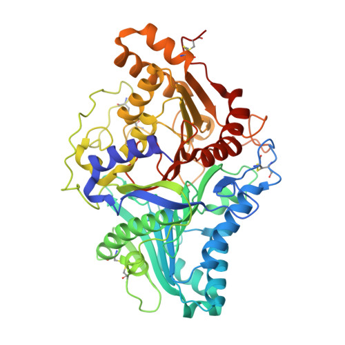 | |
UniProt | |||||
Entity Groups | |||||
| Sequence Clusters | 30% Identity50% Identity70% Identity90% Identity95% Identity100% Identity | ||||
| UniProt Group | Q53728 | ||||
Sequence AnnotationsExpand | |||||
Reference Sequence | |||||
| Ligands 1 Unique | |||||
|---|---|---|---|---|---|
| ID | Chains | Name / Formula / InChI Key | 2D Diagram | 3D Interactions | |
| MES Download:Ideal Coordinates CCD File | B [auth A] | 2-(N-MORPHOLINO)-ETHANESULFONIC ACID C6 H13 N O4 S SXGZJKUKBWWHRA-UHFFFAOYSA-N |  | ||
| Length ( Å ) | Angle ( ˚ ) |
|---|---|
| a = 61.004 | α = 90 |
| b = 85.008 | β = 90 |
| c = 99.343 | γ = 90 |
| Software Name | Purpose |
|---|---|
| REFMAC | refinement |
| Weissenberg | data collection |
| DENZO | data reduction |
| SCALEPACK | data scaling |
| MLPHARE | phasing |


