Structural Basis for the Abo Blood-Group Dependence of Plasmodium Falciparum Rosetting.
Vigan-Womas, I., Guillotte, M., Juillerat, A., Hessel, A., Raynal, B., England, P., Cohen, J.H., Bertrand, O., Peyrard, T., Bentley, G.A., Lewit-Bentley, A., Mercereau-Puijalon, O.(2012) PLoS Pathog 8: 2781
- PubMed: 22807674 Search on PubMedSearch on PubMed Central
- DOI: https://doi.org/10.1371/journal.ppat.1002781
- Primary Citation Related Structures:
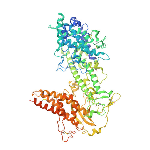
2YK0 - PubMed Abstract:
The ABO blood group influences susceptibility to severe Plasmodium falciparum malaria. Recent evidence indicates that the protective effect of group O operates by virtue of reduced rosetting of infected red blood cells (iRBCs) with uninfected RBCs. Rosetting is mediated by a subgroup of PfEMP1 adhesins, with RBC binding being assigned to the N-terminal DBL1α₁ domain. Here, we identify the ABO blood group as the main receptor for VarO rosetting, with a marked preference for group A over group B, which in turn is preferred to group O RBCs. We show that recombinant NTS-DBL1α₁ and NTS-DBL1α₁-CIDR1γ reproduce the VarO-iRBC blood group preference and document direct binding to blood group trisaccharides by surface plasmon resonance. More detailed RBC subgroup analysis showed preferred binding to group A₁, weaker binding to groups A₂ and B, and least binding to groups A(x) and O. The 2.8 Å resolution crystal structure of the PfEMP1-VarO Head region, NTS-DBL1α₁-CIDR1γ, reveals extensive contacts between the DBL1α₁ and CIDR1γ and shows that the NTS-DBL1α₁ hinge region is essential for RBC binding. Computer docking of the blood group trisaccharides and subsequent site-directed mutagenesis localized the RBC-binding site to the face opposite to the heparin-binding site of NTS-DBLα₁. RBC binding involves residues that are conserved between rosette-forming PfEMP1 adhesins, opening novel opportunities for intervention against severe malaria. By deciphering the structural basis of blood group preferences in rosetting, we provide a link between ABO blood grouppolymorphisms and rosette-forming adhesins, consistent with the selective role of falciparum malaria on human genetic makeup.
- Institut Pasteur, Unité d'Immunologie Moléculaire des Parasites, Paris, France.
Organizational Affiliation: