2WWM

Crystal structure of the Titin M10-Obscurin like 1 Ig complex in space group P1


Experimental Data Snapshot

  • Method: X-RAY DIFFRACTION
  • Resolution: 2.30 Å
  • R-Value Free: 0.252 
  • R-Value Work: 0.197 
  • R-Value Observed: 0.200 

wwPDB Validation   3D Report Full Report


This is version 1.3 of the entry. See complete history


Literature

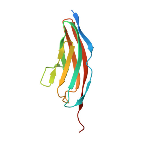
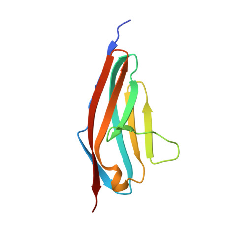
Structural Insight Into M-Band Assembly and Mechanics from the Titin-Obscurin-Like-1 Complex.

Pernigo, S.Fukuzawa, A.Bertz, M.Holt, M.Rief, M.Steiner, R.A.Gautel, M.

(2010) Proc Natl Acad Sci U S A 107: 2908

  • DOI: https://doi.org/10.1073/pnas.0913736107
  • Primary Citation of Related Structures:  
    2WP3, 2WWK, 2WWM

  • PubMed Abstract: 

    In the sarcomeric M-band, the giant ruler proteins titin and obscurin, its small homologue obscurin-like-1 (obsl1), and the myosin cross-linking protein myomesin form a ternary complex that is crucial for the function of the M-band as a mechanical link. Mutations in the last titin immunoglobulin (Ig) domain M10, which interacts with the N-terminal Ig-domains of obscurin and obsl1, lead to hereditary muscle diseases. The M10 domain is unusual not only in that it is a frequent target of disease-linked mutations, but also in that it is the only currently known muscle Ig-domain that interacts with two ligands--obscurin and obsl1--in different sarcomeric subregions. Using x-ray crystallography, we show the structural basis for titin M10 interaction with obsl1 in a novel antiparallel Ig-Ig architecture and unravel the molecular basis of titin-M10 linked myopathies. The severity of these pathologies correlates with the disruption of the titin-obsl1/obscurin complex. Conserved signature residues at the interface account for differences in affinity that direct the cellular sorting in cardiomyocytes. By engineering the interface signature residues of obsl1 to obscurin, and vice versa, their affinity for titin can be modulated similar to the native proteins. In single-molecule force-spectroscopy experiments, both complexes yield at forces of around 30 pN, much lower than those observed for the mechanically stable Z-disk complex of titin and telethonin, suggesting why even moderate weakening of the obsl1/obscurin-titin links has severe consequences for normal muscle functions.


  • Organizational Affiliation

    King's College London BHF Research Excellence Centre, Randall Division for Cell and Molecular Biophysics and Cardiovascular Division, New Hunt's House, Guy's Campus, London SE1 1UL, United Kingdom.


Macromolecules
Find similar proteins by:  (by identity cutoff)  |  3D Structure
Entity ID: 1
MoleculeChains Sequence LengthOrganismDetailsImage
OBSCURIN-LIKE PROTEIN 1A [auth C],
C [auth O]
109Homo sapiensMutation(s): 0 
UniProt & NIH Common Fund Data Resources
Find proteins for O75147 (Homo sapiens)
Explore O75147 
Go to UniProtKB:  O75147
PHAROS:  O75147
GTEx:  ENSG00000124006 
Entity Groups  
Sequence Clusters30% Identity50% Identity70% Identity90% Identity95% Identity100% Identity
UniProt GroupO75147
Sequence Annotations
Expand
  • Reference Sequence
Find similar proteins by:  (by identity cutoff)  |  3D Structure
Entity ID: 2
MoleculeChains Sequence LengthOrganismDetailsImage
TITINB [auth D],
D [auth T]
102Homo sapiensMutation(s): 0 
EC: 2.7.11.1
UniProt & NIH Common Fund Data Resources
Find proteins for Q8WZ42 (Homo sapiens)
Explore Q8WZ42 
Go to UniProtKB:  Q8WZ42
PHAROS:  Q8WZ42
GTEx:  ENSG00000155657 
Entity Groups  
Sequence Clusters30% Identity50% Identity70% Identity90% Identity95% Identity100% Identity
UniProt GroupQ8WZ42
Sequence Annotations
Expand
  • Reference Sequence
Experimental Data & Validation

Experimental Data

  • Method: X-RAY DIFFRACTION
  • Resolution: 2.30 Å
  • R-Value Free: 0.252 
  • R-Value Work: 0.197 
  • R-Value Observed: 0.200 
  • Space Group: P 1
Unit Cell:
Length ( Å )Angle ( ˚ )
a = 37.65α = 87.29
b = 44.94β = 86.91
c = 68.01γ = 68.79
Software Package:
Software NamePurpose
REFMACrefinement
MOSFLMdata reduction
SCALAdata scaling
MOLREPphasing

Structure Validation

View Full Validation Report


Entry History 

Deposition Data

Revision History  (Full details and data files)

  • Version 1.0: 2010-02-16
    Type: Initial release
  • Version 1.1: 2011-07-13
    Changes: Advisory, Version format compliance
  • Version 1.2: 2011-08-24
    Changes: Derived calculations
  • Version 1.3: 2023-12-20
    Changes: Data collection, Database references, Other, Refinement description