Evidence for Conformational Changes Upon Copper Binding to Cupriavidus Metallidurans Czce.
Petit-Haertlein, I., Girard, E., Sarret, G., Hazemann, J., Gourhant, P., Kahn, R., Coves, J.(2010) Biochemistry 49: 1913
- PubMed: 20112954 Search on PubMed
- DOI: https://doi.org/10.1021/bi100001z
- Primary Citation Related Structures:
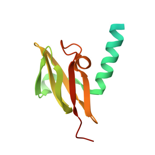
2WTO, 2WTP - PubMed Abstract:
CzcE is a periplasmic protein from Cupriavidus metallidurans CH34 that can bind four copper atoms per dimer. We have crystallized the apo form of the protein and determined its structure at 1.85 A resolution. Three Cu atoms were localized by soaking apo-CzcE crystals into a CuCl(2) solution. We identified His24 as a Cu(II) ligand in each protomer and Asp100 as a key residue for Cu binding at the interface of the dimer. The role of these amino acids was confirmed by site-directed mutagenesis and UV-visible spectroscopy. The fourth Cu atom was not located. The oxidized form of CzcE contains four Cu(II) atoms, while the reduced form contains four Cu(I) atoms. Average coordination spheres of four N or O atoms for Cu(II) and of one N or O atom and two S atoms for Cu(I) were determined by X-ray absorption spectroscopy. As there is no evidence for preformed metal-binding sites in apo-CzcE, we suggest that different conformational changes occurred upon Cu(II) or Cu(I) binding. These changes were further demonstrated by digestion experiments that gave different proteolysis patterns depending not only on the presence of the metal but also on its speciation. The ability of CzcE to bind copper and to adapt its conformation to different copper oxidation states could be related to a role in copper sensing for this protein.
- Institut de Biologie Structurale-Jean-Pierre Ebel, UMR 5075 CNRS-CEA-UJF, 41, rue Jules Horowitz, 38027 Grenoble Cedex, France.
Organizational Affiliation: